BNL’s Xiao, Yang, Liu use two beamlines to look closely at cellular structure, biochemistry
By Daniel Dunaief
Superman’s x-ray and heat vision illustrate an important problem. On the one hand, the x-ray vision comes in handy if Superman is looking outside, say, at a bank and can see thieves dressed like the Hamburgler as they try to steal from a vault. On the other hand, Superman has heat vision, which he uses in battles to blow up concrete blocks or tear open a hole in a wall.
But, aside from a few realities getting in the way, the struggle scientists using x-rays to see inside cells contend with tracks with these two abilities.
Researchers would ideally like to use x-rays to see the inner workings of a cell. X-rays can and do act like Superman’s heat vision, causing damage or destroying the cells they are trying to study.
Recently, scientists at Brookhaven National Laboratory, however, figured out how to protect and preserve cells, providing an opportunity to study them without causing damage.
Not only that, but, to extend the fictional metaphor, they used the equivalent of Wonder Twin Powers, combining the structural three-dimensional picture one beamline at the National Synchrotron Lightsource II can produce with the two-dimensional chemical image from another.
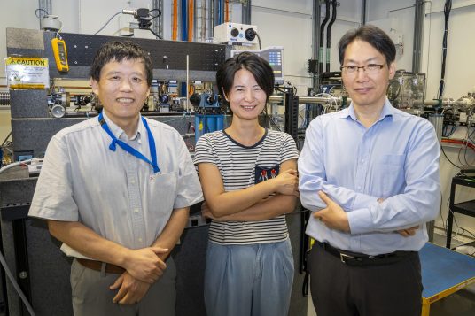
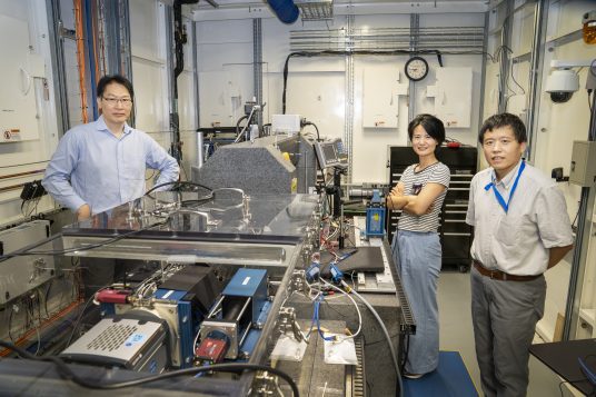
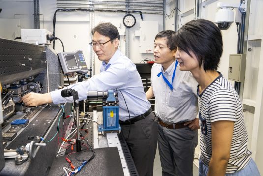
After three years of hard work, researchers including Qun Liu, structural biologist; Yang Yang, associate physicist; and Xianghui Xiao, FXI lead beamline scientist, were able to use both beamlines to create a multimodal picture of a cell on different scales and with different information.
“Each beamline can create a full picture, but providing only partial information (structure or chemicals),” Liu said. “The correlative imaging for the same cell using two different beamlines provides a more comprehensive” image.
The key to this proof of concept, Liu explained, was in developing a multi-step process to study the cells.
“The novelty is how we prepared the samples,” said Liu. “We can take the sample from one beamline, move it to a second one, and can collect data from the same orientation. Before this, it was not easy” to put together that kind of information.
In a paper published in the journal Nature Communications Biology, the scientists detailed the cell preparation technique and showcased the results.
The potential application of this technique extends in numerous directions, from finding the way new pathogens attack cells, to understanding the location and site of action of pharmacological agents, to understanding the progression of disease, among other applications.
“Our technique combines both X-ray fluorescence and X-ray nano-tomography so we can study the entire cell for both the elements and the structure correlatively,” Yang explained.
Supported by the Department of Energy Biopreparedness Initiative, the scientists are doing basic research and developing techniques and protocols and procedures in preparation for the next pandemic. They have 10 projects covering different pathogens and aspects. Liu is the principal investigator leading one of them.
To be sure, at this point, the technique for preserving and studying cells with these beamlines is in an early stage and is not available to labs, doctors, or hospitals on a routine basis to test biological samples.
Nonetheless, the approach at BNL offers an important potential direction for clinical and fundamental benefits. Clinically, it can help with disease diagnosis, while it can also be used to study stresses of cells and tissues under metal deficiency or toxicity. Many cancers include a malfunction in the homeostasis, including zinc, copper and iron.
Fixing and re-fixing
The process of preparing the samples required three steps.
The researchers started with a chemical fixation with paraformaldehyde to preserve the structure of the cell. They then used a robot that rapidly froze the sample by plunging it into liquid ethane and then transferring it to liquid nitrogen.
They freeze-dried the cells to turn the water into ice that is not crystallized. As a part of that process, they left the cells in a controlled vacuum to turn the ice slowly into gas. Removing water is key because the liquid would otherwise be too mobile for x-rays to measure anything reliably. After absorbing the x-rays, the liquid would heat up and further deform the cells.
The preparation work takes one to two days.
“If you fail in any of the steps, you have to start all over again,” said Yang.
Zihan Lin, who is a postdoctoral researcher in Liu’s lab and the first author on the paper, spent more than a year polishing and preparing the technique.
“We believe the cells were preserved [near] their close-to-native status,” said Yang.
They used an X-ray computed tomography (XCT) beamline, which provides a three-dimensional view of the structure of the cell. They also placed the samples in an X-ray fluorescence beamline (XRF), which provided a two-dimensional view of the same cells.
In the XRF beamline, scientists can find where trace elements are located inside a cell.
Liu is collaborating with researchers at other labs to understand the molecular interactions between sorghum, an important grain crop, and the fungus Colletotrichum sublineola, which can damage the leaves of the plant.
The DOE funded project is a collaboration between BNL and three other national laboratories.
Liu is grateful for the help and support he and the team received from the staff working at both beamlines, as well as from the biology department, NSLS-II, BNL, and DOE. The imaging may help create bioenergy crops with more biomass and less disease-caused yield loss, he suggested.
Future work
Current and ongoing work is focused on the potential physiological states of the cell, addressing questions such as why metals are going to specific areas.
Yang is the science lead for a team developing the Quantitative Cellular Tomography beamline at the NSLS-II. Within five years, this beamline will provide nanoscale resolution of frozen cells without requiring chemical fixation.
This beamline, which will have a light epi-fluorescence microscope, will add more detail about sub-cellular structure and will not require frozen cells to have chemical fixation.
While the proof of concept approach with these beamlines is still relatively new, Yang said she has received feedback from scientists interested in its potential.
“We have quite a few people from biology departments that are interested in this technique” to study biomass related structures, she said.
A future research direction could also involve seeing living cells. The resolution would be compromised, as the X-rays would induce changes that make it hard to separate biological processes from artifacts.
“This could be a very good research direction,” Liu added.

