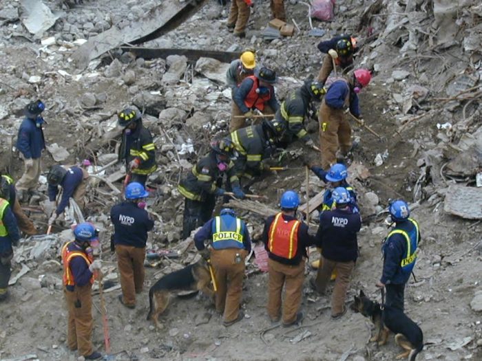
Twenty-two years after the September 11 World Trade Center attacks, responders who have suffered physical and cognitive illnesses resulting from exposures continue to be monitored by healthcare providers. Ongoing studies by investigators at the Stony Brook WTC Health and Wellness Program reveal that assessments of this patient population’s mental health and cognitive status remain on the forefront of research as we move further away from that fateful day of 9/11.
Benjamin Luft, MD, Director and Principal Investigator of the Stony Brook WTC Health and Wellness Program, and the Edmund D. Pellegrino Professor of Medicine at the Renaissance School of Medicine at Stony Brook University, and his colleagues study all aspects of responders’ health status. The program monitors approximately 13,000 WTC responders.
Previous research has shown that some responders may be experiencing cognitive difficulties earlier in life than the general population, and that PTSD, which remains one of their most common ailments, may be associated with cognitive problems and/or physical illnesses.
A compilation of new research published over the past year suggests the need to delve further into investigating the brain status of responders and their cognitive problems.
A study in the Journal of Geriatric Psychiatry and Neurology assessed more than 700 responders, many with chronic PTSD, and the relationship between having cortical atrophy and behavioral impairments. They found that individuals with PTSD start to experience more mental health symptoms as a secondary symptom to cognitive impairments. Specifically, responders with an increased risk of cortical atrophy showed behavioral impairment in motivation, mood, disinhibition, empathy and psychosis.
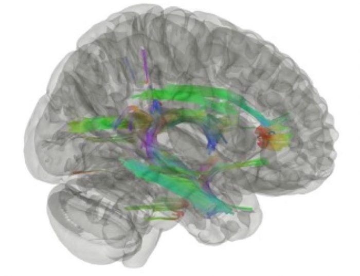
Published in Molecular Neurobiology, another study revealed that there are associations between WTC exposure duration and inflammation in the brains of responders among 99 responders who participated from 2017 to 2019, with the average age being only 56 years. Neuroinflammation was evident both in the hippocampus, a part of the brain that helps to regulate emotions and memory, and throughout much of the cerebral white matter.
A paper published in Psychological Medicine highlights research that may reveal a better way to understand responders’ PTSD symptoms, as opposed to self-reporting or screening. This work found that by using an AI program that reads the words of responders can predict their current PTSD and even the future trajectory of the illness.
Moreover, WTC investigators are developing AI programs to identify and predict psychological symptoms from facial expressions and tone of voice. AI analyzes video recordings of WTC responders. Importantly, when these methods are fully developed, they may be able to offer objective diagnostic tests for PTSD and other mental disorders.
Many responders to date have experienced mild cognitive impairment in comparison to non-responders their age.
A study that measured a key aspect of brain chemistry — proteins or biomarkers often associated with dementia and Alzheimer’s Disease — may provide specific evidence that responders need to be monitored for earlier onset dementia.
Published in the Alzheimer’s Association’s Diagnosis, Assessment and Disease Monitoring, this study illustrates that among approximately 1,000 responders — average age at 56.6 years, and some who have dementia — associations exist between WTC exposures and the prevalence of neurodegenerative proteins in their brains.
Lead author Sean Clouston, PhD, Professor in the Program of Public Health, and the Department of Family, Population, and Preventive Medicine, and colleagues found that 58 percent of responders with dementia had at least one elevated biomarker and nearly 3.5 percent had elevations in all biomarkers. The overall cohort had an increased risk of dementia associated with plasma biomarkers indicative of neurodegenerative disease.
Another core member of the Stony Brook research team, Pei-Fen Kean, PhD, Professor in the Department of Applied Mathematics and Statistics, is involved in several ongoing multi-omics research projects to help explicate pathophysiology of these disorders on molecular level and identify novel blood-based biomarkers. For example, a study in the Translational Psychiatry identified the metabolomic-proteomic signatures associated with PTSD to enhance understanding of the biological pathways implicated in PTSD.
As the collaborative work of the research teams affiliated with the Stony Brook WTC Health and Wellness Program moves forward, they will use previous findings and new methods to build their work to best assess the mental and physical health conditions of responders.


