Tags Posts tagged with "Medical Compass"

Medical Compass

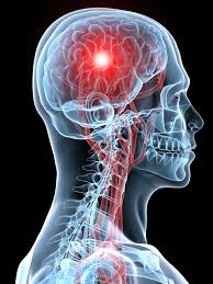
The main symptom of a heart attack is chest pain. Stock photo
As many as 35 percent of heart attacks may present without chest pain

By David Dunaief, M.D.

Dr. David Dunaief

Heart disease is the most common chronic disease in America. When we refer to heart disease, it is an umbrella term; heart attacks are one component. 

Fortunately, the incidence of heart attacks has decreased over the last several decades, as have deaths from heart attacks. However, there are still 790,000 heart attacks every year, and almost three-quarters of these are first heart attacks (1).

If you think someone is having a heart attack, call 911 as quickly as possible and have the patient chew an adult aspirin (325 mg) or four baby aspirins. While the Food & Drug Administration does not recommend aspirin for primary prevention of a heart attack, the use of aspirin here is for treatment of a potential heart attack, not prevention.

Heart attack symptoms

The main symptom is chest pain, which most people don’t have trouble recognizing. However, there are a number of other, more subtle, symptoms such as discomfort or pain in the jaw, neck, back, arms and epigastric, or upper abdominal areas. Others include nausea, shortness of breath, sweating, light-headedness and tachycardia (racing heart rate). One problem is that less than one-third of people know these other major symptoms (2). About 10 percent of patients present with atypical symptoms — without chest pain — according to one study (3).

It is not only difficult for the patient but also for the medical community, especially the emergency room, to determine who is having a heart attack. Fortunately, approximately 80 to 85 percent of chest pain sufferers are not having a heart attack. More likely, they have indigestion, reflux or other non-life-threatening ailments.

There has been a raging debate about whether men and women have different symptoms when it comes to heart attacks. Several studies speak to this topic.

Men vs. women

There is data showing that, although men have heart attacks more commonly, women are more likely to die from a heart attack (4). In a Swedish prospective (forward-looking) study, after having a heart attack, a significantly greater number of women died in hospital or near-term when compared to men. The women received reperfusion therapy, artery opening treatment that consisted of medications or invasive procedures, less often than the men.

However, recurrent heart attacks occurred at the same rate, regardless of sex. Both men and women had similar findings on an electrocardiogram; they both had what we call ST elevations. This was a study involving approximately 54,000 heart attack patients, with one-third of them being women.

One theory about why women are treated less aggressively when first presenting in the ER is that they have different and more subtle symptoms — even chest pain symptoms may be different. Women’s symptoms may include pain in the lower portion of the chest or upper portion of the abdomen and may have significantly less severe pain that could radiate or spread to the arms. But, is this true? Not according to several studies.

In one observational study, results showed that, though there were some subtle differences in chest pain, on the whole, when men and women presented with this main symptom, it was of a similar nature (5). There were 34 chest pain characteristic questions used to determine if a difference existed. These included location, quality or type of pain and duration. Of these, there was some small amount of divergence: The duration was shorter for a man (2 to 30 minutes), and pain subsided more for men than for women. The study included approximately 2,500 patients, all of whom had chest pain. The authors concluded that determination of heart attacks with chest pain symptoms should not factor in the sex of patients.

This trial involved an older population; patients were a median age of 70 for women and 59 for men, with more men having had a prior heart attack. The population difference was a conspicuous weakness of an otherwise solid study, since age and previous heart attack history are important factors.

In the GENESIS-PRAXY study, another observational study, but with a younger population, the median age of both men and women was 49. Results showed that chest pain remained the most prevalent presenting symptom in both men and women (6). However, of the patients who presented without distinct chest pain and with less specific EKG findings (non-ST elevations), significantly more were women than men. Those who did not have chest pain symptoms may have had some of the following symptoms: back discomfort; weakness; discomfort or pain in the throat, neck, right arm and/or shoulder; flushing; nausea; vomiting; and headache.

If the patients did not have chest pain, regardless of sex, the symptoms were diffuse and nonspecific. The researchers were looking at acute coronary syndrome, which encompasses heart attacks. In this case, independent risk factors for disease not related to chest pain included both tachycardia (rapid heart rate) and being female. The authors concluded that there need to be better ways to calibrate non-chest pain symptoms.

Some studies imply that as much as 35 percent of patients do not present with chest pain as their primary complaint (7).

Let’s summarize

So what have we learned about heart attack symptoms? The simplest lessons are that most patients have chest pain, and that both men and women have similar types of chest pain. However, this is where the simplicity stops and the complexity begins. The percentage of patients who present without chest pain seems to vary significantly depending on the study — ranging from less than 10 percent to 35 percent.

Non-chest pain heart attacks have a bevy of diffuse symptoms, including obscure pain, nausea, shortness of breath and light-headedness. This is seen in both men and women, although it occurs more often in women. When it comes to heart attacks, suspicion should be based on the same symptoms for both sexes. Therefore, know the symptoms, for it may be your life or a loved one’s that depends on it.

References:

(1) cdc.gov. (2) MMWR. 2008;57:175–179. (3) Chest. 2004;126:461-469. (4) Int J Cardiol. 2013;168:1041-1047. (5) JAMA Intern Med. 2014 Feb. 1;174:241-249. (6) JAMA Intern Med. 2013;173:1863-1871. (7) JAMA 2012;307:813-822.

Dr. Dunaief is a speaker, author and local lifestyle medicine physician focusing on the integration of medicine, nutrition, fitness and stress management. For further information, visit www.medicalcompassmd.com.  

Studies have shown that eating fresh fruit and cinnamon may be beneficial to diabetics. Stock photo
Fresh fruit and cinnamon may reduce risk

By David Dunaief, M.D.

Dr. David Dunaief

What causes Type 2 diabetes? It would seem like an obvious answer: obesity, right? Well, obesity is a contributing factor but not necessarily the only factor. This is important because the prevalence of diabetes is at epidemic levels in the United States, and it continues to grow. The latest statistics show that about 12.2 percent of the U.S. population aged 18 or older has Type 2 diabetes, and about 9.4 percent when factoring all ages (1).

Not only may obesity play a role, but sugar by itself, sedentary lifestyle and visceral (abdominal) fat may also contribute to the pandemic. These factors may not be mutually exclusive, of course.

We need to differentiate among sugars, because form is important. Sugar and fruit are not the same with respect to their effects on diabetes, as the research will help clarify. Sugar, processed foods and sugary drinks, such as fruit juices and soda, have a similar effect, but fresh fruit does not.

Sugar’s impact

Sugar may be sweet, but it also may be a bitter pill to swallow when it comes to its effect on the prevalence of diabetes. In an epidemiological (population-based) study, the results show that sugar may increase the prevalence of Type 2 diabetes by 1.1 percent worldwide (2). This seems like a small percentage, however, we are talking about the overall prevalence, which is around 9.4 percent in the U.S., as we noted above.

Also, the amount of sugar needed to create this result is surprisingly low. It takes about 150 calories, or one 12-ounce can of soda per day, to potentially cause this rise in diabetes. This is looking at sugar on its own merit, irrespective of obesity, lack of physical activity or overconsumption of calories. The longer people were consuming sugary foods, the higher the incidence of diabetes. So the relationship was a dose-dependent curve. Interestingly, the opposite was true as well: As sugar was less available in some countries, the risk of diabetes diminished to almost the same extent that it increased in countries where it was overconsumed.

In fact, the study highlights that certain countries, such as France, Romania and the Philippines, are struggling with the diabetes pandemic, even though they don’t have significant obesity issues. The study evaluated demographics from 175 countries, looking at 10 years’ worth of data. This may give more bite to municipal efforts to limit the availability of sugary drinks. Even steps like these may not be enough, though. Before we can draw definitive conclusion from the study, however, there need to be prospective (forward-looking) studies.

Effect of fruit

The prevailing thought has been that fruit should only be consumed in very modest amounts in patients with — or at risk for — Type 2 diabetes. A new study challenges this theory. In a randomized controlled trial, newly diagnosed diabetes patients who were given either more than two pieces of fresh fruit or fewer than two pieces had the same improvement in glucose (sugar) levels (3). Yes, you read this correctly: There was a benefit, regardless of whether the participants ate more fruit or less fruit.

This was a small trial with 63 patients over a 12-week period. The average patient was 58 and obese, with a body mass index of 32 (less than 25 is normal). The researchers monitored hemoglobin A1C (HbA1C), which provides a three-month mean percentage of sugar levels.

It is very important to emphasize that fruit juice and dried fruit were avoided. Both groups also lost a significant amount of weight while eating fruit. The authors, therefore, recommended that fresh fruit not be restricted in diabetes patients.

What about cinnamon?

It turns out that cinnamon, a spice many people love, may help to prevent, improve and reduce sugars in diabetes. In a review article, the authors discuss the importance of cinnamon as an insulin sensitizer (making the body more responsive to insulin) in animal models that have Type 2 diabetes (4).

Cinnamon may work much the same way as some medications used to treat Type 2 diabetes, such as GLP-1 (glucagon-like peptide-1) agonists. The drugs that raise GLP-1 levels are also known as incretin mimetics and include injectable drugs such as Byetta (exenatide) and Victoza (liraglutide). In a study with healthy volunteers, cinnamon raised the level of GLP-1 (5). Also, in a randomized control trial with 100 participants, 1 gram of cassia cinnamon reduced sugars significantly more than medication alone (6). The data is far too preliminary to make any comparison with FDA-approved medications. However, it would not hurt, and may even be beneficial, to consume cinnamon on a regular basis.

Sedentary lifestyle

What impact does lying down or sitting have on diabetes? Here, the risks of a sedentary lifestyle may outweigh the benefits of even vigorous exercise. In fact, in a recent study, the authors emphasize that the two are not mutually exclusive in that people, especially those at high risk for the disease, should be active throughout the day as well as exercise (7).

So in other words, the couch is “the worst deep-fried food,” as I once heard it said, but sitting at your desk all day and lying down also have negative effects. This coincides with articles I’ve written on exercise and weight loss, where I noted that people who moderately exercise and also move around much of the day are likely to lose the greatest amount of weight.

As a medical community, it is imperative that we reduce the trend of increasing prevalence by educating the population, but the onus is also on the community at large to make lifestyle changes. So America, take an active role.

References:

(1) www.cdc.gov/diabetes. (2) PLoS One. 2013;8(2):e57873. (3) Nutr J. published online March 5, 2013. (4) Am J Lifestyle Med. 2013;7(1):23-26. (5) Am J Clin Nutr. 2007;85:1552–1556. (6) J Am Board Fam Med. 2009;22:507–512. (7) Diabetologia online March 1, 2013.

Dr. Dunaief is a speaker, author and local lifestyle medicine physician focusing on the integration of medicine, nutrition, fitness and stress management. For further information, visit www.medicalcompassmd.com.      

Stock photo
Speedy diagnosis and treatment improves outcomes

By David Dunaief, M.D.

Dr. David Dunaief

TIA (transient ischemic attack) is sometimes referred to as a “mini-stroke.” This is a disservice since it makes a TIA sound like something that should be taken lightly. Ischemia is reduced or blocked blood flow to the tissue, due to a clot or narrowing of the arteries. Symptoms may last less than five minutes. However, a TIA is a warning shot across the bow that needs to be taken very seriously on its own. It may portend life-threatening or debilitating complications that can be prevented with a combination of medications and lifestyle modifications.

Is TIA common?

It is diagnosed in anywhere from 200,000 to 500,000 Americans each year (1). The operative word is “diagnosed,” because it is considered to be significantly underdiagnosed. I have helped manage patients with symptoms as understated as the onset of double vision. Other symptoms may include facial or limb weakness on one side, slurred speech or problems comprehending others, dizziness or difficulty balancing or blindness in one or both eyes (2). TIA incidence increases with age (3).

What is a TIA?

It is a brief episode of neurological dysfunction caused by focal brain ischemia or retinal ischemia (low blood flow in the back of the eye) without evidence of acute infarction (tissue death) (4). In other words, TIA has a rapid onset with potential to cause temporary muscle weakness, creating difficulty in activities such as walking, speaking and swallowing, as well as dizziness and double vision.

Though they are temporary, TIAs have potential complications, from increased risk of stroke to heightened depressive risk to even death. Despite the seriousness of TIAs, patients or caregivers often delay receiving treatment.

Stroke risk

A TIA is a stroke that lasts only a few minutes.
Stock photo

After a TIA, stroke risk goes up dramatically. Even within the first 24 hours, stroke risk can be 5 percent (5). According to one study, the incidence of stroke is 11 percent after seven days, which means that almost one in 10 people will experience a stroke after a TIA (6). Even worse, over the long term, the probability that a patient will experience a stroke reaches approximately 30 percent, one in three, after five years (7).

The EXPRESS study, a population-based study that considered the effect of urgent treatment of TIA and minor stroke on recurrent stroke, evaluated 1,287 patients, comparing their initial treatment times after experiencing a TIA or minor stroke and their subsequent outcomes (8).

The Phase 1 cohort was assessed within a median of three days of symptoms and received a first prescription within 20 days. In Phase 2, median delays for assessment and first prescription were less than one day. All patients were followed for two years after treatment. Phase 2 patients had significantly improved outcomes over the Phase 1 patients. Ninety-day stroke risk was reduced from 10 to 2 percent, an 80 percent improvement.

The study’s authors advocate for the creation of TIA clinics that are equipped to diagnose and treat TIA patients to increase the likelihood of early evaluation and treatment and decrease the likelihood of a stroke within 90 days. The moral of the story is: Treat a TIA as a stroke should be treated, the faster the diagnosis and treatment, the lower the likelihood of sequalae, or complications.

Predicting the risk of stroke

Both DWI (diffusion-weighted imaging) and ABCD2 are potentially valuable predictors of stroke after TIA. The ABCD2 is a clinical tool used by physicians. ABCD2 stands for Age, Blood pressure, Clinical features and Diabetes, and it uses a scoring system from 0 to 7 to predict the risk of a stroke within the first two days of a TIA (9).

Heart attack

In one epidemiological study, the incidence of a heart attack after a TIA increased by 200 percent (10). These were patients without known heart disease. Interestingly, the risk of heart attacks was much higher in those over 60 years of age and continued for years after the event. Just because you may not have had a heart attack within three months after a TIA, this is an insidious effect; the average time frame for patients was five years from TIA to heart attack.

Mortality

TIAs decrease overall survival by 4 percent after one year, by 13 percent after five years and by 20 percent after nine years, especially in those over age 65 (11). The reason younger patients had a better survival rate, the authors surmise, is that their comorbidity (additional diseases) profile was more favorable.

Depression

In a cohort study that involved over 5,000 participants, TIA was associated with an almost 2.5-times increased risk of depressive disorder (12). Those who had multiple TIAs had a higher likelihood of depressive disorder. Unlike with stroke, in TIA it takes much longer to diagnose depression, about three years after the event.

What can you do?

Awareness and education are important. While 67 percent of stroke patients receive education about their condition, only 35 percent of TIA patients do (13). Many risk factors are potentially modifiable, with high blood pressure being at the top of the list, as well as high cholesterol, increasing age (over 55) and diabetes.

Secondary prevention (preventing recurrence) and prevention of complications are similar to those of stroke protocols. Medications may include aspirin, antiplatelets and anticoagulants. Lifestyle modifications include a Mediterranean and DASH diet combination. Patients should not start an aspirin regimen for chronic preventive use without the guidance of a physician.

If you or someone you know has TIA symptoms, the patient needs to see a neurologist and a primary care physician and/or a cardiologist immediately for assessment and treatment to reduce risk of stroke and other long-term effects.

References:

(1) Stroke. Apr 2005;36(4):720-723; Neurology. May 13 2003;60(9):1429-1434. (2) mayoclinic.org. (3) Stroke. Apr 2005;36(4):720-723. (4) N Engl J Med. Nov 21 2002;347(21):1713-1716. (5) Neurology. 2011 Sept 27; 77:1222. (6) Lancet Neurol. Dec 2007;6(12):1063-1072. (7) Albers et al., 1999. (8) Stroke. 2008;39:2400-2401. (9) Lancet. 2007;9558;398:283-292. (10) Stroke. 2011; 42:935-940. (11) Stroke. 2012 Jan;43(1):79-85. (12) Stroke. 2011 Jul;42(7):1857-1861. (13) JAMA. 2005 Mar 23;293(12):1435.

Dr. Dunaief is a speaker, author and local lifestyle medicine physician focusing on the integration of medicine, nutrition, fitness and stress management. For further information, visit www.medicalcompassmd.com.    

Eat the colors of the rainbow to reduce the risk of dementia. Stock photo
Intensive lifestyle changes may grow protective telomeres

By David Dunaief, M.D.

Dr. David Dunaief

Dementia may be diagnosed when someone experiences loss of memory plus loss of another faculty, such as executive functioning (decision-making) or language abilities (speaking, writing or reading). The latter is known as aphasia. Alzheimer’s disease is responsible for approximately 60 to 80 percent of dementia cases (1).

Unfortunately, there are no definitive studies that show reversal or a cure for Alzheimer’s disease. This is why prevention is central to Alzheimer’s — and dementia in general.

In terms of dementia, there is good news and some disappointing news.

We will start with the good news. Though chronological age is a risk factor that cannot be changed, biological age may be adjustable. There are studies that suggest we may be able to prevent dementia through the use of both lifestyle modifications and medications.

Telomeres’ length and biological age

Biological age may be different from chronologic age, depending on a host of environmental factors that include diet, exercise and smoking. There are substances called telomeres that are found at the ends of our chromosomes. They provide stability to this genetic material. As our telomeres get shorter and shorter, our cellular aging and, ultimately, biological aging, increases.

In a preliminary case control study, dementia patients were shown to have significantly shorter telomere length than healthy patients (2). Interestingly, according to the authors, men have shorter telomere length and may be biologically older by four years than women of the same chronological age. The researchers caution that this is a preliminary finding and may not have clinical implications.

What I find most intriguing is that intensive lifestyle modifications increased telomere length in a small three-month study with patients who had low-risk prostate cancer (3). By adjusting their lifestyles, study participants were potentially able to decrease their biological ages.

Diet’s effect

Lifestyle modifications play a role in many chronic diseases and disorders. Dementia is no exception. In a prospective observational study, involving 3,790 participants, those who had the greatest compliance with a Mediterranean-type diet demonstrated a significant reduction in the risk of Alzheimer’s disease, compared to the least compliant (4). Participants were over the age of 65, demographics included substantial numbers of both black and white participants, and there was a mean follow-up of 7.6 years. Impressively, those who adhered more strictly to the diet performed cognitively as if they were three years younger, according to the authors.

Beta-carotene and vitamin C effect

In a small, preliminary case-control study (disease vs. healthy patients), higher blood levels of vitamin C and beta-carotene significantly reduced the risk of dementia, by 71 percent and 87 percent, respectively (5). The blood levels were dramatically different in those with the highest and lowest blood levels of vitamin C (74.4 vs. 28.9 µmol/L) and beta-carotene (0.8 vs. 0.2 µmol/L).

The reason for this effect may be that these nutrients help reduce oxidative stress and thus have neuroprotective effects, preventing the breakdown of neurons. This study was done in the elderly, average 78.9 years old, which is a plus, since as we age we’re more likely to be afflicted by dementia.

It is critically important to delineate the sources of vitamin C and beta-carotene in this study. These numbers came from food, not supplements. Why is this important? First, beta-carotene is part of a family of nutrients called carotenoids. There are at least 600 carotenoids in food, all of which may have benefits that are not achieved when taking beta-carotene supplements. Second, beta-carotene in supplement form may increase the risk of small-cell lung cancer in smokers (6).

Foods that contain beta-carotene include fruits and vegetables such as berries; green leafy vegetables; and orange, red or yellow vegetables like peppers, carrots and sweet potato. In my practice, I test for beta-carotene and vitamin C as a way to measure nutrient levels and track patients’ progress when they are eating a nutrient-dense diet. Interestingly, many patients achieve more than three times higher than the highest beta-carotene blood levels seen in this small study.

Impact of high blood pressure medications

For those patients who have high blood pressure, it is important to know that not all blood pressure medications are created equal. When comparing blood pressure medications in an observational study, two classes of these medications stood out. Angiotensin II receptor blockers (known as ARBs) and angiotensin-converting enzyme inhibitors (known as ACE inhibitors) reduce the risk of dementia by 53 and 24 percent, respectively, when used in combination with other blood pressure medications.

Interestingly, when ARBs were used alone, there was still a 47 percent reduction in risk; however, ACE inhibitors lost their prevention advantage. High blood pressure is a likely risk factor for dementia and can also be treated with lifestyle modifications (7). Otherwise, ARBs or ACE inhibitors may be the best choices for reducing dementia risk.

Ginkgo biloba disappoints

Ginkgo biloba, a common herbal supplement taken to help prevent dementia, may have no benefit. In the GuidAge study, ginkgo biloba was shown to be no more effective than placebo in preventing patients from progressing to Alzheimer’s disease (8). This randomized controlled trial was done in elderly patients over a five-year period with almost 3,000 participants. There was no difference seen between the treatment and placebo groups. This reinforces the results of an earlier study, Ginkgo Evaluation of Memory trial (9). Longer studies may be warranted. The authors stressed the importance of preventive measures with dementia.

You may be able to prevent dementia, whether through lifestyle modifications or, if medications are necessary, through medication selection.

References:

(1) www.uptodate.com. (2) Arch Neurol. 2012 Jul 23:1-8. (3) Lancet Oncol. 2008;9(11):1048-1057. (4) Am J Clin Nutr. 2011;93:601-607. (5) J Alzheimers Dis. 2012;31:717-724. (6) Am. J. Epidemiol. 2009; 169(7):815-828. (7) Neurology. 2005;64(2):277. (8) Lancet Neurol. 2012;11(10):851-859. (9) JAMA. 2008;300(19):2253-2262.

Dr. Dunaief is a speaker, author and local lifestyle medicine physician focusing on the integration of medicine, nutrition, fitness and stress management. For further information, visit www.medicalcompassmd.com.      

Reducing inflammation can reduce disease risk. Stock photo
C-reactive protein can be measured to identify disease risk

By David Dunaief, M.D.

One of the most widely studied biomarkers for inflammation in our bodies is high-sensitivity C-reactive protein (hsCRP), also referred to as CRP. High sensitivity means that we can measure levels as low as 0.3 mg/L more accurately.

What is the significance of the different levels? Individuals who have levels lower than 1.0 mg/L are in the optimal range for low risk for a host of diseases that are indicated by high inflammation. 

For example, with heart disease, levels of 1 to 3 mg/L represent the average risk range, and greater than 3.0 mg/L is a higher risk profile. Above 10.0 mg/L is more likely associated with other causes, such as infection and autoimmune diseases (1, 2). This biomarker is derived from the liver.

CRP is not specific to heart disease, nor is it definitive for risk of the disease. However, the upside is that it may be helpful with risk stratification, which helps us understand where we sit on a heart disease risk spectrum and with progression in other diseases, such as age-related macular degeneration, diabetic retinopathy, depression and autoimmune diseases. Let’s look at the evidence.

Stock photo

Age-related macular degeneration

Age-related macular degeneration (AMD) is the leading cause of blindness in patients over the age of 65 (3). Therefore, it is very important to help define risk stratification for this disease. In a prospective study, results showed that hsCRP levels were inversely associated with the risk of developing AMD. The group with an hsCRP greater than 3.0 mg/L had a 50 percent increased risk of developing overall AMD compared to the optimal group with hsCRP lower than 1.0 mg/L. But even more interestingly, the risk of developing neovascular, or wet, AMD increased to 89 percent in this high-risk group.

The significance of wet AMD is that it is one type of advanced-stage AMD that results in blindness. This study involved five studies where the researchers thawed baseline blood samples from middle-aged participants who had hsCRP levels measured. There were more than 2,000 participants with a follow-up as long as 20 years. According to the study’s authors, annual eye exams and lifestyle modifications, including supplements, may be able to stem this risk by reducing hsCRP.

These results reinforce those of a previous prospective study that showed that elevated hsCRP increased the risk of AMD threefold (4). This study utilized data from the Women’s Health Study, which involved over 27,000 participants. Like the study mentioned above, this one also defrosted blood samples from baseline and looked at follow-up incidence of developing AMD in initially healthy women.

The highest group had hsCRP levels over 5.2 mg/L. Additionally, when analyzing similar cutoffs for high- and low-level hsCRP, as the above trial used, those with hsCRP over 3.0 had an 82 percent increased risk of AMD compared to those with an hsCRP of lower than 1.0 mg/L.

Diabetic retinopathy

We know that diabetes affects just under 10 percent of the U.S. population and is continuing to rise. One of the complications of diabetes is diabetic retinopathy, which affects the retina (back of the eye) and is a leading cause of vision loss (5). One of the reasons for the vision loss is macular edema, or swelling, usually due to rupture of tiny blood vessels below the macula, a portion of the back of the eye responsible for central vision.

The Diabetes Control and Complications Trial (DCCT), a prospective study involving over 1,400 Type 1 diabetes patients, showed an 83 percent increased risk of developing clinically significant macular edema in the group with the highest hsCRP levels compared to those with the lowest (6). Although these results were with Type 1 diabetes, patients with Type 2 diabetes are at equal risk of diabetic retinopathy if glucose levels, or sugars, are not well controlled.

Depression

Depression is a very difficult disease to control and is a tremendous cause of disability.

Well, it turns out that inflammation is associated with depression. Specifically, in a prospective observational trial, rising levels of CRP had a linear relationship with increased risk of hospitalization due to psychological distress and depression (7). In other words, compared to levels of less than 1 mg/L, those who were 1 to 3 mg/L, 3 to 10 mg/L and greater than 10 mg/L had increased risk from 30 to 84 to 127 percent, respectively. This study involved over 70,000 patients.

How can you reduce inflammation?

This is the key question, since we now know that hsCRP is associated with systemic inflammation. In the Nurses’ Health Study, a very large, prospective observational study, the Dietary Approaches to Stop Hypertension (DASH) diet decreased the risk of both heart disease and stroke, which is impressive. The DASH diet also decreases the levels of hsCRP significantly, which was associated with a decrease in clinically meaningful end points of stroke and heart disease (8). The DASH diet is nutrient dense with an emphasis on fruits, vegetables, nuts, seeds, legumes and whole grains and a de-emphasis on processed foods, red meats, sodium and sweet beverages.

Conclusion

As the evidence shows with multiple diseases, hsCRP is a very valuable nonspecific biomarker for inflammation in the body. To stem the effects of inflammation, reducing hsCRP through lifestyle modifications and drug therapy may be a productive way of reducing risk, slowing progression and even potentially reversing some disease processes.

The DASH diet is a very powerful approach to achieving optimal levels of hsCRP without incurring potential side effects. This is a call to arms to have your levels measured, especially if you are at high risk or have chronic diseases such as heart disease, diabetes, depression and autoimmune diseases. HsCRP is a simple blood test with easy-to-obtain results.

References:

(1) uptodate.com. (2) Diabetes Technol Ther. 2006;8(1):28-36. (3) Prog Retin Eye Res. 2007 Nov;26(6):649-673. (4) Arch Ophthalmol. 2007;125(3):300-305. (5) Am J Ophthalmol. 2003;136(1):122-135. (6) JAMA Ophthalmol. 2013 Feb 7;131:1-8. (7) JAMA Psychiatry. 2013;70(2):176-184. (8) Arch Intern Med. 2008;168(7):713-720.

Dr. Dunaief is a speaker, author and local lifestyle medicine physician focusing on the integration of medicine, nutrition, fitness and stress management. For further information, visit www.medicalcompassmd.com.      

Pedometers may help achieve exercise goals

By David Dunaief, M.D.

Dr. David Dunaief

Chronic obstructive pulmonary disease, or COPD, is the third leading cause of mortality in the United States, although it’s not highlighted much in the layman’s press (1).

COPD is an umbrella term that includes emphysema, chronic bronchitis of more than three months for two consecutive years and/or chronic obstructive asthma. It is an obstructive lung disease that limits airflow. The three most common symptoms of the disease involve shortness of breath, especially on exertion, production of sputum and cough. This disease affects 6.7 percent of the U.S. population (2).

It tends to be progressive, meaning more frequent and severe exacerbations over time. Since it is a devastating and debilitating chronic disease with no cure, anything that can identify and prevent COPD exacerbations, as well as comorbidities (associated diseases), is critically important.

What are the traditional ways to reduce the risk of and treat COPD exacerbations? The most important step is to stop smoking, since 80 percent of COPD is related to smoking. Supplemental oxygen therapy and medications, such as corticosteroids, bronchodilators (beta-adrenergic agonists and anticholinergics) and antibiotics help to alleviate symptoms (3).

One of the underlying components of COPD may be chronic inflammation (4). Therefore, reducing inflammation may help to stem COPD exacerbations. There are several inflammatory biomarkers that could potentially help predict exacerbations and mortality associated with this disease, such as interleukin-6 (IL-6), C-reactive protein (CRP), leukocyte (white blood cell) count and fibrinogen (a clotting factor of the blood).

How do we reduce inflammation, which may contribute to exacerbations of this disease? Some drugs, such as statins, work partially by reducing inflammation. They may have a role in COPD. Lifestyle changes that include a high-nutrient, anti-inflammatory diet and exercise may also be beneficial.

Let’s look at the evidence.

Biomarkers for inflammation

In a population-based study with over 60,000 participants, results show that as three biomarkers (CRP, leukocyte count and fibrinogen) were elevated, the risk of COPD exacerbation increased in a linear manner (5). In other words, the risk of frequent exacerbation increased 20, 70 and 270 percent within the first year as the number of elevated biomarkers increased from one to three, compared to patients who did not have biomarker elevations.

As time progressed beyond the first year of follow-up, risk exacerbation continued to stay high. Patients with all three biomarkers elevated for longer periods had a 150 percent increased risk of frequent exacerbations. These predictions were applicable to patients with stable and with mild COPD.

In an observational study, results showed that when the biomarker IL-6 was elevated at the start of the trial in stable COPD patients, the risk of mortality increased almost 2.7-fold (6). Also, after three years, IL-6 increased significantly. Elevated IL-6 was associated with a worsening of six-minute walking distance, a parameter tied to poor physical performance in COPD patients. However, unlike the previous study, CRP did not show correlation with increased COPD exacerbation risk. This was a small trial, only involving 53 patients. Therefore, the results are preliminary.

These biomarker trials are exciting for their potential to shape treatments based on level of exacerbation risk and mortality, creating more individualized therapies. Their results need to be confirmed in a randomized controlled trial (RCT). Many of these biomarkers mentioned in the two trials are identifiable with simple blood tests at major labs.

Statin effect

Statins have been maligned for their side effects, but their efficacy has been their strong suit. An observational trial showed that statins led to at least a 30 percent reduction in the risk of COPD exacerbations, with the effect based on a dose-dependent curve (7). In other words, as the dose increased, so did the benefit.

Interestingly, even those who had taken the statin previously saw a significant reduction in COPD exacerbation risk. The duration of statin use was not important; a short use of statins, whether presently or previously, had substantial benefit. However, the greatest benefit was seen in those who had been on a medium to high dose or were on the drug currently. The researchers believe that the mechanism of action for statins in this setting has to do with their anti-inflammatory and immune-modulating effects. This was a retrospective (backward-looking) study with over 14,000 participants. We will need a prospective (forward-looking) study and an RCT to confirm the results.

Exercise

stock photo

Exercise is beneficial for almost every circumstance, and COPD is no exception. But did you know that a pedometer might improve results? In a three-month study, those with mild COPD were much more successful at achieving exercise goals and reducing exacerbations and symptoms when they used pedometers, compared to the group given advice alone (8). Pedometers gave patients objective feedback on their level of physical activity, which helped motivate them to achieve the goal of walking 9,000 steps daily. This is a relatively easy way to achieve exercise goals and reduce the risk of COPD exacerbations.

When exercising, we are told to vary our exercise routines on regular basis. One study demonstrates that this may be especially important for COPD patients (9). Results show that nonlinear periodization exercise (NLPE) training is better than traditional routines of endurance and resistance training in severe COPD patients. The goal of NLPE is to regularly alter the time spent working out, the number of sets, the number of repetitions and the intensity of the workout on a regular basis.

This study was randomized, involved 110 patients and was three months in duration. Significantly more severe COPD patients achieved their exercise goals using NLPE than the traditional approach. The group that used NLPE also had an improved quality of life response. The researchers believe that compliance with an NLPE-type program is mostly likely going to be greater because patients seem to enjoy it more.

Chronic inflammation may play a central role in COPD exacerbation. Nonspecific inflammatory biomarkers are potentially valuable for providing more personalized approach to therapy. Drugs that can control inflammation, such as statins, show promise. But don’t forget the importance of lifestyle changes, such as quitting smoking and committing to an exercise regimen that is varied and/or involves the use of a pedometer. And potentially a high-nutrient, anti-inflammatory diet will also contribute positively to reducing the frequency and severity of COPD exacerbations.

References:

(1) Natl Vital Stat Rep. 2011 Dec.;59(10):1-126. (2) cdc.gov. (3) N Engl J Med. 2002;346:988-994. (4) www.goldcopd.org. (5) JAMA. 2013;309:2353-2361. (6) Respiratory Research. 2013;14:24. (7) Am J Med. 2013 Jul;126:598-606. (8) ATS 2013 International Conference: Abstract A1360. (9) Am J Respir Crit Care Med. 2013; online Feb. 28.

Dr. Dunaief is a speaker, author and local lifestyle medicine physician focusing on the integration of medicine, nutrition, fitness and stress management. For further information, visit www.medicalcompassmd.com.    

Lowering sodium intake may have far-reaching benefits, and it is certainly achievable. Stock photo
High sodium: potassium ratio increases cardiovascular risk

By David Dunaief, M.D.

Dr. David Dunaief

We need sodium in our diets in modest amounts; however, many Americans overconsume it. Meanwhile, potassium, which we also need, is underconsumed.

More than 90 percent of people consume far too much sodium, with salt being the primary culprit (1). Sodium is found in foods that don’t even taste salty. Bread and rolls are the primary offenders. Other foods with substantial amounts of sodium are cold cuts and cured meats, cheeses, pizza (which has both bread and cheese), fresh and processed poultry, soups, meat dishes, pastas and snack foods. Foods that are processed and those prepared by restaurants are where most of our consumption occurs (2).

By contrast, only about 2 percent of people get enough potassium from their diets (3). According to one study, we would need to consume about eight sweet potatoes or 10 bananas each day to reach appropriate levels. Why is it important to reduce sodium and increase potassium? A high sodium-to-potassium ratio increases the risk of cardiovascular disease by 46 percent, according to the study, which looked at more than 12,000 Americans over almost 15 years (4). In addition, both may have significant impacts on blood pressure and cardiovascular disease.

To improve our overall health, we need to tip the sodium-to-potassium scales, consuming less sodium and more potassium. Let’s look at the evidence.

Reduced sodium

Two studies illustrate the benefits of reducing sodium in high blood pressure and normotensive (normal blood pressure) patients, ultimately preventing cardiovascular disease, including heart disease and stroke.

The first used the prestigious Cochrane review to demonstrate that blood pressure is reduced by a significant mean of −4.18 mm Hg systolic (top number) and −2.06 mm Hg diastolic (bottom number) involving both normotensive and hypertensive participants (5). When looking solely at hypertensive patients, the reduction was even greater, with a systolic blood pressure reduction of −5.39 mm Hg and a diastolic blood pressure reduction of −2.82 mm Hg.

This was a meta-analysis (a group of studies) that evaluated data from randomized clinical trials, the gold standard of studies. There were 34 trials reviewed with more than 3,200 participants. Salt was reduced from 9 to 12 grams per day to 5 to 6 grams per day. These levels were determined using 24-hour urine tests. The researchers believe there is a direct linear effect with salt reduction. In other words, the more we reduce the salt intake, the greater the effect of reducing blood pressure. The authors concluded that these effects on blood pressure will most likely result in a decrease in cardiovascular disease.

In the second study, a meta-analysis of 42 clinical trials, there was a similarly significant reduction in both systolic and diastolic blood pressures (6). This meta-analysis included adults and children. Both demographics saw a reduction in blood pressure, though the effect, not surprisingly, was greater in adults. Interestingly, an increase in sodium caused a 24 percent increased risk of stroke incidence but, more importantly, a 63 percent increased risk of stroke mortality. The risk of mortality from heart disease was increased as well, by 32 percent.

In an epidemiology modeling study, the researchers projected that either a gradual or instantaneous reduction in sodium would save lives (7). For instance, a modest 40 percent reduction over 10 years in sodium consumed could prevent 280,000 premature deaths. These are only projections, but in combination with the above studies they may be telling. The bottom line: Decrease sodium intake by almost half and increase potassium intake from foods.

Potassium’s positive effects

When we think of blood pressure, sodium comes to mind, but not enough attention is given to potassium. The typical American diet doesn’t contain enough of this mineral.

In a meta-analysis involving 32 studies, results showed that as the amount of potassium was increased, systolic blood pressure decreased significantly (8). When foods containing 3.5 to 4.7 grams of potassium were consumed, there was an impressive −7.16 mm Hg reduction in systolic blood pressure with high blood pressure patients. Anything more than this amount of potassium did not have any additional benefit. Increased potassium intake also reduced the risk of stroke by 24 percent. This effect was important.

The reduction in blood pressure was greater with increased potassium consumption than with sodium restriction, although there was no head-to-head comparison done. The good news is that potassium is easily attainable in the diet. Foods that are potassium-rich include bananas, sweet potatoes, almonds, raisins and green leafy vegetables such as Swiss chard.

Lowering sodium intake may have far-reaching benefits, and it is certainly achievable. We need to reduce our intake and give ourselves a brief period to adapt — it takes about six weeks to retrain our taste buds, once we reduce our sodium intake. We can also improve our odds by increasing our dietary potassium intake, which also has a substantial beneficial effect, striking a better sodium-to-potassium balance.

References:

(1) Am J Clin Nutr. 2012 Sep;96(3):647-657. (2) www.cdc.gov. (3) Am J Clin Nutr. 2012 Sep;96(3):647-657. (4) Arch Intern Med. 2011;171(13):1183-1191. (5) BMJ. 2013 Apr 3;346:f1325. (6) BMJ. 2013 Apr 3;346:f1326. (7) Hypertension. 2013; 61: 564-570. (8) BMJ. 2013; 346:f1378.

Dr. Dunaief is a speaker, author and local lifestyle medicine physician focusing on the integration of medicine, nutrition, fitness and stress management. For further information, visit www.medicalcompassmd.com or consult your personal physician.  

by -
0 243
Osteoarthritis is a common form of arthritis that often affects the knee. Stock photo
Lifestyle changes may slow progression
Dr. David Dunaief

Osteoarthritis is widespread. Most commonly, it affects the knees, hips and hands. There are three types of treatment: surgery, involving joint replacements of the hips or knees; medications; and nonpharmacologic approaches. The most commonly used first-line medications are acetaminophen and nonsteroidal anti-inflammatory drugs, such as ibuprofen. Unfortunately, medications mostly treat the symptoms of pain and inflammation.

However, the primary objectives in treating osteoarthritis should also include improving quality of life, slowing progression of the disease process and reducing its disabling effects (1).

Dairy and milk

When we think of dairy, specifically milk, there are two distinct camps: One believes in the benefits, and the other thinks it may contribute to disease. In this case they both may be at least partly correct. In the Osteoarthritis Initiative study, an observational study of over 2,100 patients, results showed that low-fat (1 percent) and nonfat milk may slow the progression of osteoarthritis (2). The researchers looked specifically at joint space narrowing that occurs in those with affected knee joints. Radiographic imaging changes were used at baseline and then to follow the patients for up to 12 to 48 months for changes. Compared to those who did not drink milk, patients who did saw significantly less narrowing of knee joint space.

Was it a dose-dependent response? Not necessarily. Specifically, those who drank less than three glasses/week and those who drank four to six glasses/week both saw slower progression of joint space narrowing of 0.09 mm. Seven to 10 glasses/week resulted in a 0.12 mm preservation. However, those who drank more than 10 glasses/week saw less beneficial effect, 0.06 mm preservation compared to those who did not drink milk. Interestingly, there was no benefit seen in men or with the consumption of cheese or yogurt.

However, there are significant flaws with this study. First, the patients were only asked about their dietary intake of milk at baseline; therefore their consumption could have changed during the study. Second, there was a recall bias; patients were asked to recall their weekly milk consumption for the previous 12 months before the study began. I don’t know about you, but I can’t recall my intake of specific foods for the last week, let alone for the past year. Third, there could have been confounding factors, such as orange consumption.

Oddly, this was not a dose-response curve, since the most milk consumption had less beneficial effect than lower amounts. Also, why were these effects only seen in women? Finally, researchers could not explain why low-fat or nonfat milk had this potential benefit, but cheese was detrimental and yogurt did not show benefit. We are left with more questions than answers.

Would I recommend consuming low-fat or nonfat milk? Not necessarily, but I may not dissuade osteoarthritis patients from drinking it. There are very few approaches that slow the progression of joint space narrowing.

Vitamin D

Over the last five years or so, the medical community has gone from believing that vitamin D was potentially the solution to many diseases to wondering whether, in some cases, low levels were indicative of disease, but repletion was not a change-maker. Well, in a randomized controlled trial (RCT), the gold standard of studies, vitamin D had no beneficial symptom relief, nor any disease-modifying effects (3). This two-year study of almost 150 men and women raised blood levels of vitamin D on average to 36 ng/ml, which is considered respectable. Researchers used MRI and X-rays to track their results.

Weight

This could not be an article on osteoarthritis if I did not talk about weight. In a study involving 112 obese patients, there was not only a reduction of knee symptoms in those who lost weight, but there was also disease modification, with reduction in the loss of cartilage volume around the medial tibia (4).

On the other hand, those who gained weight saw the inverse effect. A reduction of tibial cartilage is potentially associated with the need for knee replacement. The relationship was almost one-to-one; for every 1 percent of weight lost, there was a 1.2 mm³ preservation of medial tibial cartilage volume, while the exact opposite was true with weight gain.

Exercise and diet

In a study, diet and exercise trumped the effects of diet or exercise alone (5). Patients with osteoarthritis of the knee who lost at least 10 percent of their body weight experienced significant improvements in function and a 50 percent reduction in pain, as well as reduction in inflammation, compared to those who lost 5 to 10 percent and those who lost less than 5 percent. This study was a well-designed, randomized controlled single-blinded study with a duration of 18 months.

Researchers used a biomarker — IL6 — to measure inflammation. The diet and exercise group and the diet-only group lost significantly more weight than the exercise-only group, 23.3 pounds and 19.6 pounds versus 4 pounds. The diet portion consisted of a meal replacement shake for breakfast and lunch and then a vegetable-rich, low-fat dinner. Low-calorie meals replaced the shakes after six months. The exercise regimen included one hour of a combination of weight training and walking with alacrity three times per week.

Therefore, concentrate on lifestyle modifications if you want to see potentially disease-modifying effects. These include both exercise and diet. In terms of low-fat or nonfat milk, while the study had numerous flaws, if you drink milk, you might continue for the sake of osteoarthritis, but stay on the low end of consumption. And remember, the best potential effects shown are with weight loss and with a vegetable-rich diet.

References:

(1) uptodate.com. (2) Arthritis Care Res online. 2014 April 6. (3) JAMA. 2013;309:155-162. (4) Ann Rheum Dis online. 2014 Feb. 11. (5) JAMA. 2013;310:1263-1273.

Dr. Dunaief is a speaker, author and local lifestyle medicine physician focusing on the integration of medicine, nutrition, fitness and stress management. For further information, visit www.medicalcompassmd.com or consult your personal physician.     

Exercise, especially endurance-based, can reduce your risk of forming gallstones. Stock photo
Weight and inactivity are among the greatest risk factors

By David Dunaief, M.D.

Dr. David Dunaief

Gallstones affect up to 20 million Americans between the ages of 20 and 74, with a more than two times increased occurrence in women than in men, according to the NHANES III survey (1). There are two types of gallstones, 80 percent of which are cholesterol stones and 20 percent of which are pigment stones.

Common symptoms

Gallstones may be asymptomatic; however, when gallstones block either the cystic or common bile ducts, symptoms occur. Symptoms include dull or crampy abdominal pain that is exacerbated by meals and lasts one to five hours. Jaundice, which includes yellowing of skin and eyes, is another symptom. Others include nausea and vomiting, rapid heart rate, hypotension (low blood pressure) and fever (2).

Tests used for diagnosis

Blood tests include complete blood count, where there may be a rise in white blood cells; liver enzymes; and the pancreatic enzymes lipase and amylase. Diagnostic tests that have more accuracy are the endoscopic ultrasonography (EUS) and endoscopic retrograde cholangiopancreatography (ERCP); however, these are invasive. Less accurate but noninvasive tests include abdominal X-ray, ultrasound and CAT scan (CT). The tests used also depend on where the stone may be located. Hepatobiliary (HIDA) scans are accurate if the stone is located in the cystic duct. And magnetic resonance retrograde cholangiopancreatography (MRCP) is used if the stone is thought to be located in the common bile duct (2).

What are the risk factors?

There are a multitude of risk factors. Some of these are modifiable, others are not. The modifiable ones include obesity, measured by body mass index (BMI); rapid weight loss; fat consumption; hormone replacement therapy (HRT); oral contraceptives; decreased physical activity; Crohn’s disease; and certain drugs. One nonmodifiable risk factor is age; the older we get, the higher the risk, with age 40 being the demarcation line (3). Other risk factors are gender, with females being more predisposed; pregnancy; and family history (4).

Let’s look at the evidence.

Obesity risks

Obesity may play an important role. The reason obesity is implicated is potentially due to bile becoming supersaturated (5). Bile is a substance produced in the liver and stored in the gallbladder. Bile aids in the digestion or breakdown of fats in the small intestines. Crystals may form, creating cholesterol gallstones from the bile.

Body mass index

A body mass index of greater than 30 kg/m² is considered obese. In a meta-analysis of two prospective, forward-looking observational trials, Copenhagen General Population Study and the Copenhagen City Heart Study, those in the highest quintile of BMI were almost three times as likely to experience symptomatic gallstones compared to those who were in the lowest quintile (6). The highest quintile was those who had a mean BMI of 32.5 kg/m² and thus were obese, whereas those in the lowest quintile had a mean BMI of 20.9 kg/m². This is a comparison of obese to ideal BMI. Not surprisingly, since women in general have a higher risk of gallstones, they also have a higher risk when their BMI is in the obese range compared to men, a 3.36-fold increase and 1.51-fold increase, respectively.

Also, the research showed that for every 1 kg/m² increase in BMI, there was a 7 percent increase in the risk of gallstones. Those who had genetic variants that increased their likelihood of an elevated BMI had an even greater increase in gallstone risk —17 percent — per 1 kg/m². In the study population of approximately 77,000, more than 4,000 participants became symptomatic for gallstones.

Physical activity

In the Physicians’ Health Study, a prospective observational trial, those in the lowest quintile of activity between the ages of 40 and 64 had a 72 percent increased risk of gallstone formation, and those 65 and older had a 33 percent increased risk (7). Also, men who were 65 and older and watched television more than six hours a week were at least three times as likely to have gallstones as those who watched fewer hours. There was a substantial increased risk for those under 65, as well, though to a slightly lesser degree.

Diabetes rears its ugly head

Just like with obesity, diabetes is almost always a culprit for complications. In a prospective observational study, those with diabetes were at a significant 2.55 times greater risk of developing gallstones than those without (8). Again, women had a higher propensity than men, but both had significant increases in the risk of gallstone formation, 3.85 times and 2.03 times, respectively. There were almost 700 participants in this study. The researchers believe that an alteration in glucose (sugar) metabolism may create this disease risk.

Hormone replacement therapy

If you needed another reason to be leery of hormone replacement therapy (HRT), then gallstones might be it. In a prospective observational trial, women who used HRT, compared to those who did not, had a 10 percent increased risk in cholecystectomy — removal of the gallbladder — to treat gallstones (9). Though this may not sound like a large increase, oral HRT increased the risk 16 percent, and oral estrogen-only therapy without progestogens increased the risk the most, 38 percent. Transdermal HRT did not have a significantly increased risk.

It is never too early or too late to treat obesity before it causes, in this case, gallstones. With a lack of exercise, obesity is exacerbated and, not surprisingly, so is symptomatic gallstone formation. Diabetes needs to be controlled to prevent complications. HRT, unless menopausal symptoms are unbearable, continues to show why it may not be a good choice.

References:

(1) Gastroenterology. 1999;117:632. (2) emedicine.medscape.com. (3) J Hepatol. 1993;18 Suppl 1:S43. (4) uptodate.com. (5) Best Pract Res Clin Gastroenterol. 2014 Aug;28:623-635. (6) Hepatology. 2013 Dec;58:2133-2141. (7) Ann Intern Med. 1998;128:417. (8) Hepatology. 1997;2:787. (9) CMAJ. 2013;16;185:549-550.

Dr. Dunaief is a speaker, author and local lifestyle medicine physician focusing on the integration of medicine, nutrition, fitness and stress management. For further information, visit www.medicalcompassmd.com or consult your personal physician.  

Steroids can be helpful but in moderation. Stock photo
Studies suggest shorter duration treatments can be as effective, with fewer side effects

By David Dunaief, M.D.

Dr. David Dunaief

Steroids typically make headlines related to their use as a performance-enhancing drug in sports. However, if we look beyond the flashy headlines, we see that corticosteroids, or steroids, play an important role in medicine.

Medical use

Steroids have an anti-inflammatory effect. This is critical since many acute and chronic diseases are based at least partially on inflammation. Chronic diseases that benefit include allergic, inflammatory and immunological diseases (1). These types of diseases touch on almost every area of the body, from osteoarthritis and autoimmune diseases to asthma, COPD (emphysema and chronic bronchitis) and eye disorders.

Steroids are delivered orally, topically as creams, lotions and eye drops, or via injections, intravenous solutions and inhaled formulations. The most commonly known medication is prednisone, but there are many others, including prednisolone, methylprednisolone, cortisone, hydrocortisone and dexamethasone.

Their benefits can be tremendous, improving functionality and reducing pain or improving breathing. You could say they are lifesaving in some instances, and with rescue inhalers they may just be that.

The bad

However, there is a very big caveat: They come at a price. Steroids cause weight gain, increased glucose (sugars), high blood pressure, cardiovascular events, osteoporosis, change in mood (psychoses), cataracts, glaucoma, infection, peptic ulcers, Cushing’s syndrome, and the list goes on. These are among the reasons medical professionals recommend using the least amount for the shortest time.

The upshot

The good news is that a plant-based diet may have similar beneficial effects in chronic diseases as steroids without all the downsides. Let’s look at the evidence.

The role in pneumonia

Pneumonia is among the top-10 leading causes of death in the world (2). In a meta-analysis (a group of nine studies), there was no overall effect of corticosteroids in reducing the risk of mortality in community-acquired pneumonia (3). However, when the data was broken into subsets, the findings were different. In subset data of those who had severe pneumonia, there was a statistically significant 74 percent reduction in mortality. And when duration was the main focus in subgroup analysis, those who received prolonged use of steroids reduced their risk of mortality by half. 

Unfortunately, with the benefit comes an increased risk of adverse events, and this meta-analysis was no exception. There was a greater than two-times increased risk of abnormally high glucose levels with prolonged use. Thus, when giving steroids, especially for a prolonged use, it may be wise to check glucose levels.

In a randomized controlled trial (RCT), the gold standard of studies, results reinforced the beneficial effects of steroids on pneumonia. They showed that in those with both severe pneumonia and high inflammation, there was a two-thirds reduction in treatment failures when corticosteroids were added to the regimen (4). There were 120 patients involved in the study. They received antibiotics plus either methylprednisolone or placebo for five days.

Osteoarthritis: surprising results

As we know, osteoarthritis specifically of the knee is very common, and intra-articular (in the joint) injections directly into the knee are becoming routine treatment. A study compared injectable hyaluronic acid to injectable corticosteroid (5). The results showed that over three months, the corticosteroid was superior to hyaluronic acid in terms of reducing pain, 66 percent versus 43.8 percent, respectively. 

Interestingly, over the longer term, 12 months, hyaluronic acid reduced the pain and maintained its effect significantly longer than the steroid, 33 percent versus a meager 8.2 percent, respectively. Study groups received five injections of either steroid or of hyaluronic acid directly to the knee over a five-week period. Thus, steroids may not always be the most effective choice when it comes to pain reduction. Hyaluronic acid may have caused this beneficial effect by reducing inflammation, protecting cartilage and preventing cell death, according to the authors.

COPD: Length may not matter

It is not unusual to treat COPD patients with oral steroids. But what is the proper duration? The treatment paradigm has been two weeks with 40 mg of corticosteroids daily. However, results in an RCT of 600 patients showed that five days with 40 mg of corticosteroid was equivalent to 14 days of the same dosage and frequency (6). The hope is that the shorter use of steroids will mean fewer side effects. We have come a long way; prior to 1999, eight weeks of steroids was a more commonplace approach to treating acute COPD exacerbations.

Dietary effect

One of the great things about steroids is that they reduce inflammation, and we know that the basis of greater than 80 percent of chronic disease is inflammation. A plant-based diet involving lots of vegetables and fruits and some grains may have a similar effect as steroids, but without the side effects. The effect may be to modify the immune system and reduce inflammation (7).

The bioactive substances from plants thought to be involved in this process are predominantly carotenoids and the flavonoids. Thus, those patients who respond even minimally to steroids are likely to respond to a plant-based diet in much the same beneficial way without the downsides of a significant number of side effects. Diet, unlike steroids, can be used for a long duration and a high intake, with a direct relationship to improving disease outcomes.

In conclusion, it is always better to treat with the lowest effective dose for the shortest effective period when it comes to steroids. The complications of these drugs are enumerable and must always be weighed against the benefits. Sometimes, other drugs may have more beneficial effects over the long term, such as hyaluronic acid injections for knee osteoarthritis. A plant-based diet, with anti-inflammatory properties similar to steroids, may be a useful alternative for chronic disease or may be used alongside these drugs, possibly reducing their dosage and duration.

References:

(1) uptodate.com. (2) N Engl J Med. 1995;333(24):1618-1624. (3) PLoS One. 2012;7(10):e47926. (4) JAMA. 2015;313(7):677-686. (5) Open Access Rheum 2015;7:9-18. (6) JAMA. 2013;309(21):2223-2231. (7) Int J Vitam Nutr Res. 2008 Dec;78(6):293-298.

Dr. Dunaief is a speaker, author and local lifestyle medicine physician focusing on the integration of medicine, nutrition, fitness and stress management. For further information, visit www.medicalcompassmd.com or consult your personal physician.