Tags Posts tagged with "Dr. David Dunaief"

Dr. David Dunaief

Deer ticks are widely known as the sole carrier of the bacteria that causes Lyme disease in humans. Stock photo
The lingering effects of Lyme can be debilitating

By David Dunaief, M.D.

Dr. David Dunaief

After a spring where we’ve spent more than our fair share of time indoors, summer’s heat is finally here. Many of us are taking advantage of the weather to enjoy day hikes and picnics along the shoreline or bike rides through wooded areas.

The summer’s heat also means that tick season is in full swing. This means we need to be aware of Borrelia burgdorferi, better known as the bacterium that causes Lyme disease. This bacterium is typically found in the deer tick, also known as the blacklegged tick.

What do deer ticks look like? They are small and can be as tiny as a pencil tip or the size of a period at the end of a sentence. The CDC.gov site is a great resource for tick images and other information related to Lyme disease.

If you have been bitten by a tick, you should remove it with forceps, tweezers or protected fingers (paper) as close to the skin as possible and pull slow and steady straight up. Do not crush or squeeze the tick; doing so may spread infectious disease (1). In a study, petroleum jelly, fingernail polish, a hot kitchen match and 70 percent isopropyl alcohol all failed to properly remove a tick. The National Institutes of Health recommend not removing a tick with oil (2).

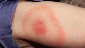
When a tick is removed within 36 to 48 hours, the risk of infection is quite low (3). However, a patient can be given a prophylactic dose of the antibiotic doxycycline, one dose of 200 mg, if a bulls-eye rash — a red outer ring and red spot in the center — has not occurred, and it is within 72 hours of tick removal (4). Those who took doxycycline had significantly lower risk of developing the bulls-eye rash and thus Lyme disease; however, treatment with doxycycline did sometimes cause nausea.

Lyme Symptoms

Stock photo

There are three stages of Lyme disease: early stage, where the bacteria are localized; early disseminated disease, where the bacteria have spread throughout the body; and late stage disseminated disease. Symptoms for early localized stage and early disseminated disease include the bulls-eye rash, which occurs in about 80 percent of patients, with or without systemic symptoms of fatigue (54 percent), muscle pain and joint pain (44 percent), headache (42 percent), neck stiffness (35 percent), swollen glands (23 percent) and fever (16 percent) (5).

Early disseminated disease may cause neurological symptoms such as meningitis, cranial neuropathy (Bell’s palsy) and motor or sensory radiculoneuropathy (nerve roots of spinal cord). Late disseminated disease can cause Lyme arthritis (inflammation in the joints), heart problems, facial paralysis, impaired memory, numbness, pain and decreased concentration (2).

Lyme carditis is a rare complication affecting 1.1 percent of those with disseminated disease, but it can result in sudden cardiac death (6). If there are symptoms of chest pain, palpitations, light-headedness, shortness of breath or fainting, clinicians should suspect Lyme carditis.

Preventing Lyme

According to the Centers for Disease Control and Prevention, we should wear protective clothing, spray ourselves with insect repellent that includes at least 20 percent DEET and treat our yards (4). Always check your skin and hair for ticks after walking through a woody or tall grassy area. Many of us on Long Island have ticks in the yard, so remember to check your pets; even if treated, they can carry ticks into the house.

Diagnosing Lyme

Lyme disease often can be diagnosed within the clinical setting or with a blood test. When it comes to serologic or blood tests, the CDC recommends an ELISA test followed by a confirmatory Western blot test (3). However, testing immediately after being bitten by a tick is not useful, since the test will tend to be negative, regardless of infection or not (4). It takes about one to two weeks for IgM antibodies to appear and two to six weeks for IgG antibodies (5). These antibodies sometimes remain elevated even after successful treatment with antibiotics.

Does chronic Lyme disease exist?

There has been a debate about whether there is something called “chronic Lyme” disease. The research, unfortunately, has not shown consistent results that indicate that it exists. In one analysis, the authors note that the definition of chronic Lyme disease is obfuscated and that extended durations of antibiotics do not prevent or alleviate post-Lyme syndromes, according to several prospective trials (7).

The authors do recognize that there are prolonged neurologic symptoms in a subset population that may be debilitating even after the treatment of Lyme disease. These authors also suggest that there may be post-Lyme disease syndromes with joint pain, muscle pain, neck and back pain, fatigue and cognitive impairment.

Ultimately, the IDSA (Infectious Diseases Society of America) argues in favor of recognizing post-Lyme disease syndromes, while the ILADS (International Lyme and Associated Diseases Society) believes chronic Lyme exists.

Regardless, the lingering effects of Lyme can be debilitating. This may be as a result of systemic inflammation (8). Systemic inflammation and its symptoms can be improved significantly with dietary and other lifestyle modifications.

The CDC recommends that physicians look beyond Lyme for other possible diagnoses before diagnosing someone with chronic Lyme disease (9).

Prevention is key to helping stem Lyme disease. If this is not possible, treating prophylactically when pulling off a tick is an important step. Contact your physician as soon as you notice a tick. If you have a bulls-eye rash and it is early, then treatment of antibiotics for two to three weeks needs to be started right away. If it is prolonged and disseminated, then treatment should be for approximately three to four weeks with antibiotics. If it has affected the central nervous system, then IV antibiotics could be needed.

References:

(1) Pediatrics. 1985;75(6):997. (2) nlm.nih.gov. (3) cdc.gov. (4) Clin Infect Dis. 2008;47(2):188. (5) uptodate.com. (6) MMWR. 2014;63(43):982-983. (7) Expert Rev Anti Infect Ther. 2011;9(7):787-797. (8) J Infect Dis. 2009;199(9:1379-1388). (9) JAMA Intern Med. online Nov. 3, 2014.

Dr. David Dunaief is a speaker, author and local lifestyle medicine physician focusing on the integration of medicine, nutrition, fitness and stress management. For further information, visit www.medicalcompassmd.com.

Consuming four or more servings of legumes per week has shown to reduce the risk of heart disease. Stock photo
Even small dietary changes move us closer to being ‘heart attack proof’

By David Dunaief, M.D.

Dr. David Dunaief

We can significantly reduce the occurrence of heart disease, the number one killer in the United States, by making modest lifestyle changes.

Heart disease is a term that captures a number of disorders, from coronary artery disease, which can cause heart attacks, to valve issues and heart failure, which is a problem with the pumping mechanism. Here, our focus will be on coronary artery disease and their resulting heart attacks.

According to the Centers for Disease Control and Prevention, there are 805,000 heart attacks in the U.S.  annually, and 200,000 of these occur in those who’ve already had a first heart attack (1). Here, I will provide specifics on how to make changes to protect you and your family, regardless of family history.

The evidence continues to highlight lifestyle changes, including diet, as the most important factors in preventing heart disease. Changes that garner a big bang for your buck include the consumption of chocolate, legumes, nuts, fiber and omega-3 polyunsaturated fatty acids (PUFAs).

Treat yourself – cocoa’s benefits

Preliminary evidence shows that two pieces of chocolate a week may decrease the risk of a heart attack by 37 percent, compared to those who consume less (2). However, the authors warned against the idea that more is better. In fact, high fat and sugar content and calorically dense aspects may have detrimental effects when consumed at much higher levels. There is a fine line between potential benefit and harm. The benefits may be attributed to micronutrients referred to as flavonols.

I usually recommend that patients have one to two squares – about one-fifth to two-fifths of an ounce – of high-cocoa-content dark chocolate daily. Who says prevention has to be painful?

Increase your fiber intake

Fiber has a dose-response relationship to reducing risk. In other words, the more fiber intake, the greater the reduction in risk. In a meta-analysis of 10 studies, results showed for every 10-gram increase in fiber, there was a corresponding 14 percent reduction in the risk of a cardiovascular event and a 27 percent reduction in the risk of heart disease mortality (3). The authors analyzed data that included over 90,000 men and 200,000 women.

The average American consumes about 16 grams per day of fiber (4). The Academy of Nutrition and Dietetics recommends 14 grams of fiber for each 1,000 calories consumed, or roughly 25 grams for women and 38 grams for men (5). Therefore, we can significantly reduce our risk of heart disease if we increase our consumption of fiber to reach the recommended levels. Good sources of fiber are fruits and vegetables with the edible skin or peel, beans and lentils, and whole grains.

Legumes’ impact

In a prospective (forward-looking) cohort study, the First National Health and Nutrition Examination Survey Epidemiologic Follow-up Study (NHEFS), legumes reduced the risk of coronary heart disease by a significant 22 percent. Those who consumed four or more servings per week, compared to those who consumed less than one serving, saw this effect. The legumes used in this study included beans, peas and peanuts (6). There were over 9,500 men and women involved, spanning 19 years of follow-up.

I recommend that patients consume at least one to two servings a day, or 7 to 14 a week. Imagine the impact that could have, compared to the modest four servings per week used to reach statistical significance in this study.

A nutty solution

In a study with over 45,000 men, there were significant reductions in coronary heart disease with omega-3 polyunsaturated fatty acids (PUFAs). Both plant-based and seafood-based omega-3s showed these effects (7). Good sources of omega-3s from plant-based sources include nuts, such as walnuts, and ground flaxseed.

Your ultimate goal should be to become “heart attack proof,” a term used by Dr. Sanjay Gupta and reinforced by Dr. Dean Ornish. Ideally, this requires a plant-based diet. But even modest changes in diet will result in significant risk reductions. The more significant the lifestyle changes you make, the closer you will come to achieving this goal.

References:

(1) cdc.gov. (2) BMJ 2011; 343:d4488. (3) Arch Intern Med. 2004 Feb 23;164(4):370-376. (4) NHANES 2009-2010 Data Brief No. 12. Sep 2014. (5) eatright.org. (6) Arch Intern Med. 2001 Nov 26;161(21):2573-2578. (7) Circulation. 2005 Jan 18;111(2):157-164.

Dr. David Dunaief is a speaker, author and local lifestyle medicine physician focusing on the integration of medicine, nutrition, fitness and stress management. For further information, visit www.medicalcompassmd.com.

Stock photo
Early diagnosis is crucial to treatment success

By David Dunaief, M.D.

Dr. David Dunaief

Diabetic retinopathy is an umbrella term for microvascular complications of diabetes that can lead to blurred vision and blindness. There are at least three different disorders that comprise it: dot and blot hemorrhages, proliferative diabetic retinopathy and diabetic macular edema. The latter two are the ones most likely to cause vision loss. Our focus for this article will be on diabetic retinopathy as a whole and on diabetic macular edema, more specifically.

Diabetic retinopathy is the number one cause of vision loss in those who are 25 to 74 years old (1). Risk factors include duration of diabetes, glucose (sugar) that is not well-controlled, smoking, high blood pressure, kidney disease, pregnancy and high cholesterol (2).

What is diabetic macula edema, also referred to as DME? Its signature is swelling caused by extracellular fluid accumulating in the macula (3). The macula is the region of the eye with greatest visual acuity. A yellowish oval spot in the central portion of the retina — in the inner segment of the back of the eye —it is sensitive to light. When fluid builds up from leaking blood vessels, there is potential for vision loss.

Those with the longest duration of diabetes have the greatest risk of DME (4). Unfortunately, many patients are diagnosed with DME after it has already caused vision loss. If not treated early, patients can experience permanent loss of vision (5). Herein lies the challenge.

In a cross-sectional study (a type of observational study) using NHANES data from 2005-2008, among patients with DME, only 45 percent were told by a physician that diabetes had affected their eyes (6). Approximately 46 percent of patients reported that they had not been to a diabetic nurse educator, nutritionist or dietician in more than a year — or never.

The problem is that the symptoms of vision loss don’t necessarily occur until the latter stages of the disorder. According to the authors, there needs to be an awareness campaign about the importance of getting your eyes examined on an annual basis if you have diabetes. Many patients are unaware of the association between vision loss and diabetes.

Treatment options                                             

While DME is traditionally treated with lasers, intravitreal (intraocular — within the eye) injections of a medication known as ranibizumab (Lucentis) may be as effective.

The results from a randomized controlled trial, the gold standard of studies, showed that intravitreal (delivery directly into the eye) injections with ranibizumab, whether given prompt laser treatments or treatments delayed for at least 24 weeks, were equally effective in treating DME (7).

Increased risk with diabetes drugs

You would think that drugs to treat type 2 diabetes would prevent DME from occurring as well. However, in the THIN trial, a retrospective (backward-looking) study, a class of diabetes drugs, thiazolidinediones, which includes Avandia and Actos, actually increased the occurrence of DME compared to those who did not use these oral medications (8). Those receiving these drugs had a 1.3 percent incidence of DME at year one, whereas those who did not had a 0.2 percent incidence. This incidence was persistent through the 10 years of follow-up. [Note that DME is not the only side effect of these drugs. There are important FDA warnings of other significant issues.]

To make matters worse, those who received both thiazolidinediones and insulin had an even greater incidence of DME. There were 103,000 diabetes patients reviewed in this trial. It was unclear whether the drugs, because they were second-line treatments, or the severity of the diabetes itself may have caused these findings.

This is in contrast to a previous ACCORD eye sub-study, a cross-sectional analysis, which did not show an association between thiazolidinediones and DME (9). This study involved review of 3,473 participants who had photographs taken of the fundus (the back of the eye).

What does this ultimately mean? Both of these studies were not without weaknesses. It was not clear how long the patients had been using the thiazolidinediones in either study or whether their sugars were controlled and to what degree. The researchers were also unable to control for all other possible confounding factors (10). Thus, there needs to be a prospective (forward-looking) trial done to sort out these results.

Diet

The risk of progression of diabetic retinopathy was significantly lower with intensive blood sugar controls using medications, one of the few positive highlights of the ACCORD trial (11). Medication-induced intensive blood sugar control also resulted in increased mortality and no significant change in cardiovascular events. But an inference can be made: A nutrient-dense, plant-based diet that intensively controls blood sugar is likely to decrease the risk of diabetic retinopathy complications (12, 13).

The best way to avoid diabetic retinopathy is obviously to prevent diabetes. Barring that, it’s to have sugars well-controlled. If you or someone you know has diabetes, it is imperative that they get a yearly eye exam from an ophthalmologist so that diabetic retinopathy is detected as early as possible, before permanent vision loss occurs. It is especially important for those diabetes patients who are taking the oral diabetes class thiazolidinediones.

References:

(1) Diabetes Care. 2014;37 (Supplement 1):S14-S80. (2) JAMA. 2010;304:649-656. (3) www.uptodate.com. (4) JAMA Ophthalmol online. 2014 Aug. 14. (5) www.aao.org/ppp. (6) JAMA Ophthalmol. 2014;132:168-173. (7) ASRS. Presented 2014 Aug. 11. (8) Arch Intern Med. 2012;172:1005-1011. (9) Arch Ophthalmol. 2010 March;128:312-318. (10) Arch Intern Med. 2012;172:1011-1013. (11) www.nei.nih.gov. (12) OJPM. 2012;2:364-371. (13) Am J Clin Nutr. 2009;89:1588S-1596S.

Dr. David Dunaief is a speaker, author and local lifestyle medicine physician focusing on the integration of medicine, nutrition, fitness and stress management. For further information, visit www.medicalcompassmd.com. 

GERD is a common condition in which the esophagus becomes irritated or inflamed because of acid backing up from the stomach. Stock photo
You may avoid medications by making simple changes

By David Dunaief, M.D.

Dr. David Dunaief

Wherever you look there is an advertisement for the treatment of heartburn or indigestion, both of which are related to reflux disease.

Reflux typically results in symptoms of heartburn and regurgitation, with stomach contents going backward up the esophagus. For some reason, the lower esophageal sphincter, the valve between the stomach and esophagus, inappropriately relaxes. No one is quite sure why it happens with some people and not others. Of course, a portion of reflux is physiologic (normal functioning), especially after a meal (1). As such, it typically doesn’t require medical treatment.

Gastroesophageal reflux disease (GERD), on the other hand, differs in that it’s long-lasting and more serious, affecting as much as 28 percent of the U.S. population (2). Can you understand why pharmaceutical firms give it so much attention?

GERD risk factors are diverse. They range from lifestyle — obesity, smoking cigarettes and diet — to medications, like calcium channel blockers and antihistamines. Other medical conditions, like hiatal hernia and pregnancy, also contribute (3). Diet issues include triggers like spicy foods, peppermint, fried foods and chocolate.

Smoking and Salt

One study showed that both smoking and salt consumption added to the risk of GERD significantly (4). Risk increased 70 percent in people who smoked. Surprisingly, people who used table salt regularly saw the same increased risk as seen with smokers.

Medications

The most common and effective medications for the treatment of GERD are H2 receptor blockers (e.g., Zantac and Tagamet), which partially block acid production, and proton pump inhibitors (e.g., Nexium and Prevacid), which almost completely block acid production (5). Both classes of medicines have two levels: over-the-counter and prescription strength. Here, I will focus on PPIs, for which more than 100 million prescriptions are written every year in the U.S. (6).

The most frequently prescribed PPIs include Prilosec (omeprazole), Protonix (pantoprazole), Nexium (esomeprazole), and Prevacid (lansoprazole). They have demonstrated efficacy for short-term use in the treatment of Helicobacter pylori-induced (bacteria overgrowth in the gut) peptic ulcers, GERD symptoms and complication prevention and gastric ulcer prophylaxis associated with NSAID use (aspirin, ibuprofen, etc.) as well as upper gastrointestinal bleeds.

However, they are often used long-term as maintenance therapy for GERD. PPIs used to be considered to have mild side effects. Unfortunately, evidence is showing that this may not be true. Most of the data in the package inserts is based on short-term studies lasting weeks, not years. The landmark study supporting long-term use approval was only one year, not 10 years. However, maintenance therapy usually continues over many years.

Side effects that have occurred after years of use are increased risk of bone fractures and calcium malabsorption; Clostridium difficile, a bacterial infection in the intestines; potential vitamin B12 deficiencies; and weight gain (7).

Bacterial infection risks

The FDA warned that patients who use PPIs may be at increased risk of a bacterial infection called C. difficile. This is a serious infection that occurs in the intestines and requires treatment with antibiotics. Unfortunately, it only responds to a few antibiotics and that number is dwindling. In the FDA’s meta-analysis, 23 of 28 studies showed increased risk of infection. Patients need to contact their physicians if they develop diarrhea when taking PPIs and the diarrhea doesn’t improve (8).

B12 deficiencies

Suppressing hydrochloric acid produced in the stomach may result in malabsorption issues if turned off for long periods of time. In a study where PPIs were associated with B12 malabsorption, it usually took at least three years’ duration to cause this effect. B12 was not absorbed properly from food, but the PPIs did not affect B12 levels from supplementation (9). Therefore, if you are taking a PPI chronically, it is worth getting your B12 and methylmalonic acid (a metabolite of B12) levels checked and discussing possible supplementation with your physician if you have a deficiency.

Lifestyle modifications

A number of modifications can improve GERD, such as raising the head of the bed about six inches, not eating prior to bedtime and obesity treatment, to name a few (10). In the same study already mentioned with smoking and salt, fiber and exercise both had the opposite effect, reducing the risk of GERD (5). This was a prospective (forward-looking) trial. The analysis by Journal Watch suggests that the fiber effect may be due to its ability to reduce nitric oxide production, a relaxant for the lower esophageal sphincter (11).

Obesity

In one study, obesity exacerbated GERD. What was interesting about the study is that researchers used manometry, which measures pressure, to show that obesity increases the pressure on the lower esophageal sphincter significantly (12). Intragastric (within the stomach) pressures were higher in both overweight and obese patients on inspiration and on expiration, compared to those with normal body mass index. This is yet another reason to lose weight.

Eating close to bedtime       

Though it may be simple, it is one of the most powerful modifications we can make to avoid GERD. A study that showed a 700 percent increased risk of GERD for those who ate within three hours of bedtime, compared to those who ate four hours or more prior to bedtime. Of note, this is 10 times the increased risk of the smoking effect (13). Therefore, it is best to not eat right before bed and to avoid “midnight snacks.”

Although there are a number of ways to treat GERD, the most comprehensive have to do with modifiable risk factors. Drugs have their place in the arsenal of choices, but lifestyle changes are the first — and most effective — approach in many instances. Consult your physician before stopping PPIs, since there may be rebound hyperacidity (high acid produced) if they are stopped abruptly.

References:

(1) Gastroenterol Clin North Am. 1996;25(1):75. (2) Gut. 2014 Jun; 63(6):871-80. (3) emedicinehealth.com. (4) Gut 2004 Dec.; 53:1730-1735. (5) Gastroenterology. 2008;135(4):1392. (6) Proton Pump Inhibitor, ClinCalc DrugStats Database, Version 20.0. Updated December 23, 2019. Accessed June 23, 2020. (7) World J Gastroenterol. 2009;15(38):4794–4798. (8) www.FDA.gov/safety/medwatch/safetyinformation. (9) Linus Pauling Institute; lpi.oregonstate.edu. (10) Arch Intern Med. 2006;166:965-971. (11) JWatch Gastro. Feb. 16, 2005. (12) Gastroenterology 2006 Mar.; 130:639-649. (13) Am J Gastroenterol. 2005 Dec.;100(12):2633-2636.

Dr. David Dunaief is a speaker, author and local lifestyle medicine physician focusing on the integration of medicine, nutrition, fitness and stress management. For further information, visit www.medicalcompassmd.com. 

Dairy may not be as beneficial as we have been led to believe. Stock Photo
Does calcium really reduce risk?

By David Dunaief, M.D.

Dr. David Dunaief

The prevalence of osteoporosis and low bone mass increase dramatically as we age. According to the Centers for Disease Control, over 48 percent of those ages 65 and older in the U.S. are affected by low bone mass, and 16.4 percent by osteoporosis (1).

Why do we care? Because they may lead to increased risk of fracture and, subsequently, lower mobility, which may have significant quality of life impacts (2). That is what we know. But what about what we think we know?

For decades we have been told that if we want strong bones, we need to consume dairy. This has been drilled into our brains since we were toddlers. Dairy has calcium and is fortified with vitamin D, so it could only be helpful, right? Not necessarily.

The data is mixed, but studies indicate that dairy may not be as beneficial as we have been led to believe. Even worse, it may be harmful. The operative word here is “may.” We will investigate this further. Vitamin D and calcium are good for us. But do supplements help prevent osteoporosis and subsequent fractures? Again, the data are mixed, but supplements may not be the answer for those who are not deficient.

Holes in the dairy paradigm

The results of a large, observational study involving men and women in Sweden showed that milk may be harmful (3). When comparing those who consumed three or more cups of milk daily to those who consumed less than one, there was a 93 percent increased risk of mortality in women between the ages of 39 and 74. There was also an indication of increased mortality based on dosage.

For every one glass of milk consumed there was a 15 percent increased risk of death in these women. There was a much smaller, but significant, 3 percent per glass increased risk of death in men. Women experienced a small, but significant, increased risk of hip fracture, but no increased risk in overall fracture risk. There was no increased risk of fracture in men, but there was no benefit either. There were higher levels of biomarkers that indicate oxidative stress and inflammation found in the urine.

This study was 20 years in duration and is eye-opening. We cannot make any decisive conclusions, only associations, since it is not a randomized controlled trial. But it does get you thinking. The researchers surmise that milk has high levels of D-galactose, a simple sugar that may increase inflammation and ultimately contribute to this potentially negative effect, whereas other foods have many-fold lower levels of this substance.

Ironically, the USDA recommends that, from 9 years of age through adulthood, we consume up to three servings of dairy per day (4). This is interesting, since the results from the previous study showed the negative effects at this recommended level of milk consumption. The USDA may want to rethink these guidelines.

Prior studies show milk may not be beneficial for preventing osteoporotic fractures. Specifically, in a meta-analysis that used data from the Nurses’ Health Study for women and the Health Professionals Follow-up Study for men, neither men nor women saw any benefit from milk consumption in preventing hip fractures (5).

Calcium disappointments

Unfortunately, it is not only milk that may not be beneficial. In a meta-analysis involving a group of observational studies, there was no statistically significant improvement in hip fracture risk in those men or women ingesting at least 300 mg of calcium from supplements and/or food on a daily basis (6).

The researchers did not differentiate the types of foods containing calcium. In a group of randomized controlled trials analyzed in the same study, those taking 800 to 1,600 mg of calcium supplements per day also saw no increased benefit in reducing nonvertebral fractures. In fact, in four clinical trials the researchers actually saw an increase in hip fractures among those who took calcium supplements. A weakness of the large multivaried meta-analyses is that vitamin D baseline levels, exercise and phosphate levels were not considered.

Vitamin D benefit

Finally, though the data is not always consistent for vitamin D, when it comes to fracture prevention, it appears it may be valuable. In a meta-analysis involving 11 randomized controlled trials, vitamin D supplementation resulted in a reduction in fractures (7). When patients were given a median dose of 800 IUs (ranging from 792 to 2,000 IUs) of vitamin D daily, there was a significant 14 percent reduction in nonvertebral fractures and an even greater 30 percent reduction in hip fractures in those 65 years and over. However, vitamin D in lower levels showed no significant ability to reduce fracture risk.

Just because something in medicine is a paradigm does not mean it’s correct. Milk may be an example of this. No definitive statement can be made about calcium, although even in randomized controlled trials with supplements, there seemed to be no significant benefit. Of course, the patients in these trials were not necessarily deficient in calcium or vitamin D.

In order to get benefit from vitamin D supplementation to prevent fracture, patients may need at least 800 IUs per day, which is the Institute of Medicine’s recommended amount for a relatively similar population as in the study.

Remember that studies, though imperfect, are better than tradition alone. Prevention and treatment therefore should be individualized, and deficiency in vitamin D or calcium should usually be treated, of course. Please, talk to your doctor before adding or changing any supplements.

References:

(1) cdc.gov (2) JAMA. 2001;285:785-795. (3) BMJ 2014;349:g6015. (4) health.gov. (5) JAMA Pediatr. 2014;168(1):54-60. (6) Am J Clin Nutr. 2007 Dec;86(6):1780-1790. (7) N Engl J Med. 2012 Aug. 2;367(5):481.

Dr. David Dunaief is a speaker, author and local lifestyle medicine physician focusing on the integration of medicine, nutrition, fitness and stress management. For further information, visit www.medicalcompassmd.com. 

Over 30 million people in the United States suffer from some form of eczema. Stock Photo
Most treatments focus on managing symptoms of itchiness or redness

By David Dunaief, M.D

Dr. David Dunaief

Referred to more broadly as atopic dermatitis, the cause of eczema is unknown, but it is thought that nature and nurture are both at play (1). Eczema is a chronic inflammatory process that involves symptoms of pruritus (itching) pain, rashes and erythema (redness) (2).

Eczema is common in both children and adults. In the United States, it’s estimated that over seven percent of the adult population is afflicted (3), with twice as many females as males affected (4). Ranging in severity from mild to moderate to severe, adults tend to have moderate to severe eczema.

Treatments for eczema run the gamut from over-the-counter creams and lotions to prescription steroid creams to systemic (oral) steroids and injectable biologics. Some use phototherapy for severe cases, but the research on phototherapy is scant. Antihistamines are sometimes used to treat the itchiness. Also, lifestyle modifications may play an important role, specifically diet. Two separate studies have shown an association between eczema and fracture, which we will investigate further.

Let’s look at the evidence.

Eczema isn’t just superficial

Eczema may also be related to broken bones, according to several studies. For example, one observational study showed that those with eczema had a 44 percent increased risk of injury causing limitation and an even more disturbing 67 percent risk of bone fracture and bone or joint injury for those 30 years and older (5). And if you have both fatigue or insomnia and eczema, you are at higher risk for bone or joint injury than having one or the other alone. One reason for increased fracture risk, the researchers postulate, is the use of corticosteroids in treatment.

Steroids may weaken bone, ligaments and tendons and may cause osteoporosis by decreasing bone mineral density. Chronic inflammation may also contribute to the risk of bone loss. There were 34,500 patients involved in the study, ranging in age from 18 to 85. For those who have eczema and have been treated with steroids, it may be wise to have a DEXA (bone) scan.

Supplements’ effectiveness

The thought of supplements somehow seems more appealing for some than medicine. There are two well-known supplements for helping to reduce inflammation, evening primrose oil and borage oil. Are these supplements a good replacement for – or addition to – medications? The research is really mixed, leaning toward ineffective.

In a meta-analysis (involving seven randomized controlled trials, the gold standard of studies), evening primrose oil was no better than placebo in treating eczema (6). The researchers also looked at eight studies of borage oil and found there was no difference from placebo in terms of symptom relief. One positive is that these supplements only had minor side effects. But don’t look to supplements for significant help.

Injectable solutions

Dupilumab is a biologic monoclonal antibody (7). In trials, this injectable drug showed good results, improving outcomes for moderate to severe eczema sufferers when topical steroids alone were not effective. Like any drug therapy, it is not without side effects, though.

Topical probiotics

When we think of probiotics, we think of taking a pill. However, there are also potentially topical probiotics with atopic dermatitis. In preliminary in-vitro (in a test tube) studies, the results look intriguing and show that topical probiotics from the human microbiome (gut) could potentially work as well as steroids (8). This may be part of the road to treatments of the future. However, this is in very early stage of development.

What about lifestyle modifications?

In a Japanese study involving over 700 pregnant women and their offspring, results showed that when the women ate either a diet high in green and yellow vegetables, beta carotene or citrus fruit there was a significant reduction in the risk of the child having eczema of 59 percent, 48 percent and 47 percent, respectively, when comparing highest to lowest consumption quartiles (9).

Elimination diets may also play a role. One study’s results showed when eggs were removed from the diet in those who were allergic, according to IgE testing, eczema improved significantly (10).

From an anecdotal perspective, I have seen very good results when treating patients who have eczema with dietary changes. My patient population includes about 15 to 20 percent of patients who suffer some level of eczema. For example, a young adult had eczema mostly on the extremities. When I first met the patient, these were angry, excoriated, erythematous and scratched lesions. However, after several months of a vegetable-rich diet, the patient’s skin had all but cleared.

I also have a personal interest in eczema. I suffered from hand eczema, where my hands would become painful and blotchy and then crack and bleed. This all stopped for me when I altered my diet many years ago.

Eczema exists on a spectrum from annoying to significantly affecting a patient’s quality of life (11). Supplements may not be the solution, at least not borage oil or evening primrose oil. However, there may be promising topical probiotics ahead and medications for the hard to treat. It might be best to avoid long-term systemic steroid use; it could not only impact the skin but also may impact the bone. Lifestyle modifications appear to be very effective, at least at the anecdotal level.

References:

(1) Acta Derm Venereol (Stockh) 1985;117 (Suppl.):1-59. (2) uptodate.com. (3) J Inv Dermatol. 2017;137(1):26-30. (4) BMC Dermatol. 2013;13(14). (5) JAMA Dermatol. 2015;151(1):33-41. (6) Cochrane Database Syst Rev. 2013;4:CD004416. (7) Medscape.com. (8) ACAAI 2014: Abstracts P328 and P329. (9) Allergy. 2010 Jun 1;65(6):758-765. (10) J Am Acad Dermatol. 2004;50(3):391-404. (11) Contact Dermatitis 2008; 59:43-47.

Dr. David Dunaief is a speaker, author and local lifestyle medicine physician focusing on the integration of medicine, nutrition, fitness and stress management. For further information, visit www.medicalcompassmd.com. 


As a general goal, aim for at least 30 minutes of moderate physical activity every day. METRO photo
Low inflammation may play a significant role in healthy aging

By David Dunaief, M.D.

Dr. David Dunaief

What does it mean to you to “grow old gracefully?” For people I speak with, it means to be independent, mobile, mentally alert, and not burdened by chronic illnesses. In other words, to maintain a good quality of life.

The number of 90-year-olds is growing by leaps and bounds. According to the National Institutes of Health, those who were more than 90 years old increased by 2.5 times over a 30-year period from 1980 to 2010 (1). This group is among what researchers refer to as the “oldest-old,” which includes those aged 85 and older.

What do these people have in common? According to one study, they tend to have fewer chronic morbidities or diseases. Thus, they tend to have a better quality of life with a greater physical functioning and mental acuity (2).

In a study of centenarians, genetics played a significant role. Characteristics of this group were that they tended to be healthy and then die rapidly, without prolonged suffering (3).

Factors that predict one’s ability to reach this exclusive club may involve both genetics and life-style choices. Let’s look at the research.

Exercise

We are told time and time again to exercise. But how much do we need, and how can we get the best quality? In a 2014 study, the results showed that 5 to 10 minutes of daily running, regardless of the pace, can have a significant impact on life span by decreasing cardiovascular mortality and all-cause mortality (4).

Amazingly, even if participants ran fewer than six miles per week at a pace slower than 10-minute miles, and even if they ran only one to two days a week, there was still a decrease in mortality compared to nonrunners. Here is the kicker: Those who ran for this very short amount of time potentially added three years to their life span. There were 55,137 participants ranging in age from 18 to 100 years old.

An accompanying editorial to this study noted that more than 50 percent of people in the United States do not meet the current recommendation of at least 30 minutes of moderate exercise per day (5).

Diet

A long-standing paradigm has been that we need to eat sufficient animal protein. However, cracks have developed in this theory, especially as it relates to longevity. In an observational study using NHANES III data, results show that those who ate a high-protein diet (greater than 20 percent from protein) had a twofold increased risk of all-cause mortality, a four-times increased risk of cancer mortality and a four-times increased risk of dying from diabetes (6). This was over a considerable duration of 18 years and involved almost 7,000 participants ranging in age at the start of the study from 50 to 65.

However, this did not hold true if the protein source was plants. In fact, a high-protein plant diet may reduce the risks, not increase them. The reason, according to the authors, is that animal protein may increase insulin growth factor-1 and growth hormones that have detrimental effects on the body.

Interestingly, those who are over age 65 may benefit from more animal protein in reducing the risk of cancer. However, there was a significantly increased risk of diabetes mortality across all age groups eating a high animal protein diet. The researchers therefore concluded that lower animal protein may be wise at least during middle age.

The Adventists Health Study 2 trial reinforced this data. It looked at Seventh-day Adventists, a group that emphasizes a plant-based diet, and found that those who ate animal protein up to once a week had a significantly reduced risk of dying over the next six years compared to those who were more frequent meat eaters (7). This was an observational trial with over 73,000 participants and a median age of 57 years old.

Inflammation

In the Whitehall II study, a specific marker for inflammation was measured, interleukin-6. The study showed that higher levels did not bode well for participants’ longevity (8). In fact, if participants had elevated IL-6 (>2.0 ng/L) at both baseline and at the end of the 10-year follow-up period, their probability of healthy aging decreased by almost half.

The takeaway from this study is that IL-6 is a relatively common biomarker for inflammation that can be measured with a simple blood test offered by most major laboratories. This study involved 3,044 participants over the age of 35 who did not have a stroke, heart attack or cancer at the beginning of the study.

The bottom line is that, although genetics are important for longevity, so too are lifestyle choices. A small amount of exercise, specifically running, can lead to a substantial increase in healthy life span. 

Protein from plants may trump protein from animal sources in reducing the risk of mortality from all causes, from diabetes and from heart disease. This does not necessarily mean that one needs to be a vegetarian to see the benefits. IL-6 may be a useful marker for inflammation, which could help predict healthy or unhealthy outcomes. Therefore, why not have a discussion with your doctor about testing to see if you have an elevated IL-6? Lifestyle modifications may be able to reduce these levels.

References:

(1) nia.nih.gov. (2) J Am Geriatr Soc. 2009;57:432-440. (3) Future of Genomic Medicine (FoGM) VII. Presented March 7, 2014. (4) J Am Coll Cardiol. 2014;64:472-481. (5) J Am Coll Cardiol. 2014;64:482-484. (6) Cell Metab. 2014;19:407-417. (7) JAMA Intern Med. 2013;173:1230-1238. (8) CMAJ. 2013;185:E763-E770.

Dr. David Dunaief is a speaker, author and local lifestyle medicine physician focusing on the integration of medicine, nutrition, fitness and stress management. For further information, visit www.medicalcompassmd.com. 

Studies show that modest wine consumption may reduce cardiovascular risks. METRO photo

By David Dunaief, M.D.

Dr. David Dunaief

By now, we have all likely heard that soda – with 39 grams of sugar per 12-ounce can – is associated with an increased risk of diabetes. Bur did you know that wine has a very low amount of sugar: about 1.2 grams of sugar in a five-ounce serving? Even more surprising, it may have benefits in reducing complications associated with diabetes.

Why is this important? The current rate of diabetes among the U.S. adult population is 12.2 percent, while another 84 million U.S. adults have prediabetes (HbA1C of 5.7-6.4 percent) (1).

For those with diabetes, cardiovascular risk and severity may not be equal between the sexes. In two trials, women with type 2 diabetes had greater cardiovascular risk than men. In one retrospective study, women with diabetes were hospitalized due to heart attacks at a more significant rate than men, though both had substantial increases in risk, 162 percent and 96 percent, respectively (2).

What may reduce risks of disease and/or complications? Fortunately, we are not without options. These include timing of blood pressure medications, lifestyle modifications (diet and exercise) and, yes, wine.

Diet vs. metformin for prevention

All too often in the medical community, we are guilty of reaching for drugs and either overlooking lifestyle modifications or expecting that patients will fail with them. This is not only disappointing, but it is a disservice; lifestyle changes may be more effective in preventing this disease. In a head-to-head comparison study (Diabetes Prevention Program), diet plus exercise bests metformin for diabetes prevention (3). This study was performed over 15 years of duration in 2,776 participants who were at high risk for diabetes because they were overweight or obese and had elevated sugars.

There were three groups in the study: those receiving a low-fat, low-calorie diet with 15 minutes of moderate cardiovascular exercise; those taking metformin 875 mg twice a day; and a placebo group. Diet and exercise reduced the risk of diabetes by 27 percent, while metformin reduced it by 18 percent over the placebo, both reaching statistical significance. Note that, while these are impressive results that speak to the use of lifestyle modification and to metformin, this is not an optimal diabetes diet.

I’ll drink to that!

Alcohol in general has mixed results. Wine is no exception. However, the CASCADE trial, a randomized controlled trial, considered the gold standard of studies, shows wine may have heart benefits in well-controlled patients with type 2 diabetes by altering the lipid (cholesterol) profile (4).

Patients were randomized into three groups, each receiving a drink with dinner nightly; one group received five ounces of red wine, another five ounces of white wine, and the control group drank five ounces of water. Those who drank the red wine saw a significant increase in their “good cholesterol” HDL levels, an increase in apolipoprotein A1 (the primary component in HDL) and a decrease in the ratio of total cholesterol-to-HDL levels compared to the water-drinking control arm. In other words, there were significant beneficial cardiometabolic changes.

White wine also had beneficial cardiometabolic effects, but not as great as red wine. However, white wine did improve glycemic (sugar) control significantly compared to water, whereas red wine did not. Also, slow metabolizers of alcohol in a combined red and white wine group analysis had better glycemic control than those who drank water. This study had a two-year duration and involved 224 patients. All participants were instructed to follow a Mediterranean-type diet.

Does this mean diabetes patients should start drinking wine? Not necessarily, because this is a small, though well-designed, study. Wine does have calories, and these were also well-controlled type 2 diabetes patients who generally were nondrinkers.

Blood pressure medications’ surprising results

Interestingly, taking blood pressure medications at night has an odd benefit, lowering the risk of diabetes (5). In a study, there was a 57 percent reduction in the risk of developing diabetes in those who took blood pressure medications at night rather than in the morning.

It seems that controlling sleep-time blood pressure is more predictive of risk for diabetes than morning or 48-hour ambulatory blood pressure monitoring. This study had a long duration of almost six years with about 2,000 participants.

The blood pressure medications used in the trial were ACE inhibitors, angiotensin receptor blockers (ARBs) and beta blockers. The first two medications have their effect on the renin-angiotensin-aldosterone system (RAAS) of the kidneys. According to the researchers, the drugs that blocked RAAS in the kidneys had the most powerful effect on preventing diabetes. Furthermore, when sleep systolic (top number) blood pressure was elevated one standard deviation above the mean, there was a 30 percent increased risk of type 2 diabetes. Interestingly, the RAAS-blocking drugs are the same drugs that protect kidney function when patients have diabetes.

We need to reverse the trend toward higher diabetes prevalence. Diet and exercise are the first line for prevention. Even a good, but nonideal, diet had better results, in comparison to medication. A modest amount of wine, especially red, may have effects that reduce cardiovascular risk. Blood pressure medications taken at night, especially those that block RAAS in the kidneys, may help significantly to prevent diabetes.

References:

(1) cdc.gov. (4) Journal of Diabetes and Its Complications 2015;29(5):713-717. (3) Lancet Diabetes Endocrinol. Online Sept. 11, 2015. (4) Ann Intern Med. 2015;163(8):569-579. (5) Diabetologia. Online Sept. 23, 2015.

Dr. David Dunaief is a speaker, author and local lifestyle medicine physician focusing on the integration of medicine, nutrition, fitness and stress management. For further information, visit www.medicalcompassmd.com. 

Studies have shown 50 percent fewer cardiac events with CoQ10 supplementation. Stock photo
Supplementation may improve outcomes

By David Dunaief, M.D.

Dr. David Dunaief

Heart attacks and heart disease get a lot of attention, but chronic heart failure is often overlooked by the press. The reason may be that heart failure is not acute like a heart attack.

To clarify by using an analogy, a heart attack is like a tidal wave whereas heart failure is like a tsunami. You don’t know it’s coming until it may be too late. Heart failure is an insidious (slowly developing) disease and thus may take years before it becomes symptomatic. It also increases the risk of heart attack and death.

There are about 6.5 million Americans with heart failure (1), and heart failure contributed to one in eight deaths in 2017 (2).

Heart failure (HF) occurs when the heart’s pumping is not able to keep up with the body’s demands and may decompensate. There are two types — systolic and diastolic. The basic difference is that the ejection fraction, the output of blood with each contraction of the left ventricle of the heart, is more or less preserved in diastolic HF, while it can be significantly reduced in systolic HF.

We have more evidence-based medicine, or medical research, on systolic heart failure. Fortunately, both types can be diagnosed with the help of an echocardiogram, an ultrasound of the heart. The signs and symptoms may be similar, as well, and include shortness of breath on exertion or when lying down, edema or swelling, reduced exercise tolerance, weakness and fatigue. Major risk factors for heart failure include diabetes, coronary artery disease, high blood pressure, obesity, smoking, poor diet, being sedentary and drinking alcohol excessively.

Typically, heart failure is treated with blood pressure medications, such as beta blockers, ACE inhibitors and angiotensin receptor blockers. We are going to look at how diet, iron and the supplement CoQ10 impact heart failure.

Effect of diet

If we look beyond the usual risk factors mentioned above, oxidative stress may play an important role as a contributor to HF. Oxidative stress is thought to potentially result in damage to the inner lining of the blood vessels, or endothelium, oxidation of cholesterol molecules and a decrease in nitric oxide, which helps vasodilate blood vessels.

In a population-based, prospective (forward-looking) study, called the Swedish Mammography Cohort, results show that a diet rich in antioxidants reduces the risk of developing HF (3). In the group that consumed the most nutrient-dense foods, there was a significant 42 percent reduction in the development of HF, compared to the group that consumed the least. According to the authors, the antioxidants were derived mainly from fruits, vegetables, whole grains, coffee and chocolate. Fruits and vegetables were responsible for the majority of the effect.

This nutrient-dense approach to diet increased oxygen radical absorption capacity. Oxygen radicals have been implicated in cellular damage and DNA damage, potentially as a result of increasing chronic inflammation. What makes this study so impressive is that it is the first of its kind to investigate antioxidants from the diet and their impacts on heart failure prevention.

This was a large study, involving 33,713 women, with good duration — follow-up was 11.3 years. There are limitations to this study, since it is an observational study, and the population involved only women. Still, the results are very exciting, and it is unlikely there is a downside to applying this approach to the population at large.

CoQ10 supplementation

Coenzyme Q10 is a substance produced by the body that helps the mitochondria (the powerhouse of the cell) produce energy. It is thought of as an antioxidant. 

Results of the Q-SYMBIO study, a randomized controlled trial, showed an almost 50 percent reduction in the risk of all-cause mortality and 50 percent fewer cardiac events with CoQ10 supplementation (4). This one randomized controlled trial followed 420 patients for two years who had severe heart failure. This involved using 100 mg of CoQ10 three times a day compared to placebo.

The lead author goes as far as to suggest that CoQ10 should be part of the paradigm of treatment. This the first new “drug” in over a decade to show survival benefits in heart failure.

A meta-analysis involving 13 studies of CoQ10 supplementation with HF confirmed that CoQ10 resulted in ejection fraction improvements among patients with less severe stages of HF, although the authors suggest that studies with more diverse demographics and that refine and compare dose responses are warranted (5).

Thus, if you have heart failure, you may want to discuss CoQ10 with your doctor.

Iron deficiency

Anemia and iron deficiency are not synonymous, since iron deficiency can occur without anemia. A recent observational study that followed 753 heart failure patients for almost two years showed that iron deficiency without anemia increased the risk of mortality in heart failure patients by 42 percent (6).

In this study, iron deficiency was defined as a ferritin level less than 100 μg/L (the storage of iron) or, alternately, transferrin saturation less than 20 percent (the transport of iron) with a ferritin level in the range 100–299 μg/L.

The authors conclude that iron deficiency is potentially more predictive of clinical outcomes than anemia, contributes to the severity of HF and is common in these patients. Thus, it behooves us to try to prevent heart failure through dietary changes, including high levels of antioxidants, because it is not easy to reverse the disease. Those with HF should have their ferritin and iron levels checked, for these are correctable. 

I am not typically a supplement advocate; however, based on the latest results, CoQ10 seems like a compelling therapy to reduce risk of further complications and potentially death. Consult with your doctor before taking CoQ10 or any other supplements, especially if you have heart failure.

References:

(1) Circulation. 2019;139(10):e56–528. (2) cdc.gov. (3) Am J Med. 2013 Jun:126(6):494-500. (4) JACC Heart Fail. 2014 Dec;2(6):641-649. (5) Am J Clin Nutr. 2013 Feb; 97(2): 268–275. (6) Am Heart J. 2013;165(4):575-582.

Dr. David Dunaief is a speaker, author and local lifestyle medicine physician focusing on the integration of medicine, nutrition, fitness and stress management. For further information, visit www.medicalcompassmd.com. 

To reduce binge eating, take the dog for a walk while social distancing. METRO photo

By David Dunaief, M.D.

Dr. David Dunaief

Obesity is an ongoing struggle for many in the United States. The latest statistics suggest that 40 percent of the population is obese. Obesity is a disease unto itself and is defined by a BMI (body mass index) of >30 kg/m2, but obesity can also be defined by excess body fat, which is more important than BMI.

Obesity has been associated with COVID-19, especially in the U.S. In a study involving 5700 hospitalized COVID-19 patients in the NYC area, the most common comorbidities were obesity, high blood pressure and diabetes (1). Of those who were hospitalized, 41.7 percent were obese.

In a study in China, results showed that those who were overweight were 86 percent more likely to have severe COVID-19 pneumonia, and that percentage increased to 142 percent when obesity is reached (2). The study has yet to be peer-reviewed, but it complements other studies.

Another study from France indicates that those with a BMI >35 (severely obese), were more likely to be put on ventilators (3).

In fact, one study’s authors suggested quarantining should be longer in obese patients because of the potential for prolonged viral shedding compared to those in the normal range for weight (4). And though age is a risk factor for COVID-19, among those younger than 60 and obese, there is a two-times increased risk of being admitted to the hospital, according to a 3,615-patient study at NYU Langone Health (5).

Why are you at higher risk for severe COVID-19 with obesity? 

According to the prevailing theory, obesity may interfere with mechanical aspects of breathing, thus increasing airway resistance and make gas in exchange more difficult in the lung. It may also impede on lung volume by exerting pressure on the lungs and may involve weaker muscles necessary for respiration (6).

Why is excess fat more important than BMI? 

First, some who have elevated BMI may not have a significant amount of fat; they may actually have more innate muscle. More than 25 percent of my patient population is “solidly built,” which means they have greater muscle mass as well as too much excess fat. (I have a body analysis scale that detects muscle mass and fat through two different currents of ohms.) Visceral fat is the most important, since it’s the fat that lines the organs, including the lungs.

For another, fat cells have adipokines, specific cell communicators found in fat cells that communicate with other fat cells but also other systems such as the brain, immune system, muscles, and liver. Adipokines can be mediators of both inflammation and insulin resistance, according to an en-docrinology study (7). In a study of over 4,000 patients with COVID-19, the author suggests that inflammation among obese patients may be an exacerbating factor for hospitalizations and severe illness (8). 

If we defined obesity as being outside the normal fat range – normal ranges are roughly 11-22 per-cent for men and 22-34 percent for women – then close to 70 percent of Americans are “obese.”

Inflammation reduction and weight-loss combined

In a randomized controlled trial with 75 participants comparing a plant-based diet to a control diet, there was a greater than 14 lbs. weight reduction and roughly 10 lbs. fat reduction over a 14-week period (9). Of the weight lost, about 70 percent was excess fat. Remember, excess body fat, through adipokines, may be inflammatory and increase the risk of severe COVID-19. 

The weight reduction with a plant-based approach may involve the increase in fiber, reduction in dietary fat and increased burning of calories after the meal, according to Physician’s Committee for Responsible Medicine (PCRM) (10).

You also want a diet that has been shown to reduce inflammation.

We are currently submitting a small study for publication involving 16 patients from my clinical practice. It shows that those who ate a whole food plant-based LIFE (low inflammatory foods every-day) diet over a seven-day period had a significant decrease in inflammation measured by hsCRP (high sensitivity c-reactive protein). This occurred in those who completely changed their diets to the LIFE diet, but also occurred in those who simply added a greens and fruit-based smoothie daily to their existing diet.

In my practice, I have seen a number of patients lose a substantial amount of weight, but also excess body fat, over a short period. For instance, a 70-year-old male lost 19 lbs. of weight and 12 lbs. of excess body fat over a six-week period. His inflammation, which was very high to start, dropped substantially to the border of optimal levels, using hsCRP as the inflammation measurement. This patient and many others have seen tandem reductions in both weight and inflammation. To boot, this was a cardiac patient whose cardiologist had considered a stent, but later said he did not need it after reducing his inflammation.

Exercise to reduce binge eating

While sheltering in place with fewer physical activities available, it is very tempting to binge eat or use food as a leisure activity. But there is a way around this. 

In patients who are overweight and obese, those who exercised compared to those who were sedentary, showed a significant reduction in binge eating over a 12-week intervention (11). The participants at baseline had a mean BMI of 30.6 kg/m2 and a mean age of 43 years. Of the 46 participants, almost two-thirds were women. Exercise can be as easy as walking or running outside while social distancing; doing exercises with your own body weight, such as calisthenics; taking online exercise classes (of which there are plenty); or using exercise equipment you have at home, might help allay binge eating.

If COVID-19 does not convince you that losing excess body fat is important, then consider that obesity contributes to, or is associated with, many other chronic diseases like cardiovascular disease, high blood pressure, and high cholesterol, which also contribute to severe COVID-19. Thus, there is an imperative to lose excess body fat. Now, while we’re sheltering in place, is the time to work on it.

References:

(1) JAMA. online April 22, 2020. (2) https://doi.org/10.2139/ssrn.3556658 (2020). (3) Obesity. online April 9, 2020. (4) Acta Diabetol. 2020 Apr 5: 1–6. (5) Clin Infect Dis. Online April 9, 2020. (6) Chron. Respir. Dis. 5, 233–242 (2008). (7) Front Endocrinol (Lausanne). 2013; 4:71. (8) MedRxiv.com. (9) Nutr Diabetes. 2018; 8: 58. (10) Inter Journal of Disease Reversal and Prevention 2019;1:1. (11) Med Sci Sports Exerc. 2020;52(4):900-908.

Dr. Dunaief is a speaker, author and local lifestyle medicine physician focusing on the integration of medicine, nutrition, fitness and stress management. For further information, visit www.medicalcompassmd.com.