Tags Posts tagged with "Cold Spring Harbor Laboratory"

Cold Spring Harbor Laboratory

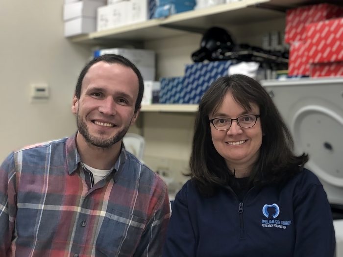
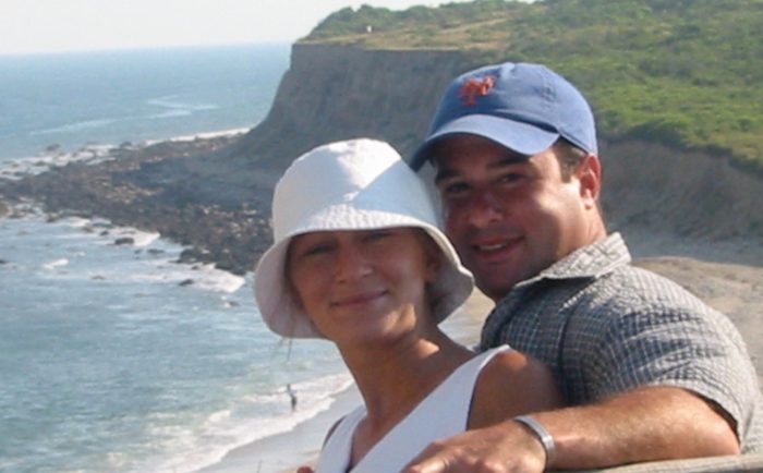
Michael Schatz and Aspyn Palatnick. Photo by Lauryl Palatnick

By Daniel Dunaief

Michael Schatz, Adjunct Associate Professor at Cold Spring Harbor Laboratory, saw some similarities to his own life when he met the then 14-year old Aspyn Palatnick.

Palatnick, who was a student at Cold Spring Harbor High School, had been developing games for the iPhone. When he was that age, Schatz, who is also a Bloomberg Distinguished Associate Professor of Computer Science and Biology at Johns Hopkins University, stayed up late into the evening programming his home computer and building new software systems.

Meeting Palatnick eight years ago was a “really special happenstance,” Schatz said. He was “super impressed” with his would-be young apprentice.

When he first met Schatz, Palatnick explained in an email that he “realized early on that he would be an invaluable mentor across research, computer science, and innovation.”

Palatnick was looking for the opportunity to apply some of the skills he had developed in making about 10 iPhone games, including a turtle racing game, to real-world problems.

Knowing that Palatnick had no formal training in computer science or genetics, Schatz spent the first several years at the white board, teaching him core ideas and algorithms.

“I was teaching him out of graduate student lecture notes,” Schatz said.

Schatz and Palatnick, who graduated with a bachelors and master’s from the University of Pennsylvania and works at Facebook, have produced a device which they liken to a “tricorder” from Star Trek. Using a smart phone or other portable technology, the free app they created called iGenomics is a mobile genome sequence analyzer.

The iPhone app complements sequencing devices Oxford Nanopore manufactures. A mobile genetic sequencer not only could help ecologists in the field who are studying the genetic codes for a wide range of organisms, but it could also be used in areas like public health to study the specific gene sequences of viruses like SARS-CoV-2, which causes COVID-19.

In a paper published in GigaScience, Schatz and Palatnick describe how to use iGenomics to study flu genomes extracted from patients. They also have a tutorial on how to use iGenomics for COVID-19 research.

While developing the mobile sequencing device wasn’t the primary focus of Schatz’s work, he said he and others across numerous departments at Johns Hopkins University spent considerable time on it this summer, as an increasing number of people around the world contracted the virus.

“It very rapidly became how I was spending the majority of my time,” said Schatz.

Palatnick is pleased with the finished product.

“We’ve made DNA sequence analysis portable for the first time,” he explained in an email.

Palatnick said the app had to use the same algorithms as traditional genomics software running on supercomputers to ensure that iGenomics was accurate and practical. Building algorithms capable of rendering DNA alignments and mutations as users tapped, scrolled and pinched the views presented a technical hurdle, Palatnick wrote.

While Schatz is optimistic about the vaccinations that health care workers are now receiving, he said a mass vaccination program introduces new pressure on the virus.

“We and everyone else are watching with great interest to see if [the vaccinations] cause the virus to mutate,” Schatz said. “That’s the big fear.”

Working with the sequences from Nanopore technology, iGenomics can compare the entire genome to known problematic sequences quickly. Users need to get the data off the Oxford Nanopore device and onto the app. They can do that using email, from Dropbox or the web. 

In prior viral outbreaks, epidemiologists traveled with heavier equipment to places like West Africa to monitor the genome of Ebola or to South and Central America to study the Zika virus genome.

“There’s clearly a strong need to have this capability,” Schatz said.

Another iGenomics feature is that it allows users to airdrop any information to people, even when they don’t have internet access.

Schatz urged users to ensure that they use a cloud-based system with strong privacy policies before considering such approaches, particularly with proprietary data or information for which privacy is critical.

As for COVID-19, people with the disease have shown enough viral mutations that researchers can say whether the strain originated in Europe or China.

“It’s kind of like spelling mistakes,” Schatz said. “There are enough spelling mistakes where [researchers] could know where it came from.”

Palatnick described iGenomics as an “impactful” tool because the app has increased the population of people who can explore the genome from institutional researchers to anyone with an iPhone or iPad.

In the bigger picture, Schatz is broadly interested in learning how the genome creates differences.

“It’s important to understand these messages for the foods we eat, the fuels we use, the medicines we take,” Schatz said. “The next frontier is all about interpretation. One of the most powerful techniques is comparing one genome to another.”

Schatz seeks out collaborators in a range of fields and at numerous institutions, including Cold Spring Harbor Laboratory.

Schatz and W. Richard McCombie, Professor at CSHL, are studying the genomes of living fossils. These are species that haven’t evolved much over millions of years. They are focusing on ancient trees in Australia that have, more or less, the same genetic make up they did 100 million years ago.

As for Palatnick, Schatz described his former intern and tricorder creating partner as a “superstar in every way.” Schatz said it takes considerable fortitude in science, in part because it takes years to go from an initial idea on a napkin to something real.

Down the road, Schatz wouldn’t be surprised if Palatnick took what he learned and developed and contributed to the founding of the next Twitter or Facebook.

“He has that kind of personality,” Schatz said.

Dennis Plenker Photo by Bob Giglione, 2020/ CSHL

By Daniel Dunaief

If the job is too easy, Dennis Plenker isn’t interested.

He’s found the right place, as the research investigator in Cold Spring Harbor Laboratory Cancer Center Director Dave Tuveson’s lab is tackling pancreatic cancer, one of the more intractable forms of cancer.

Plenker joined Tuveson’s lab in 2017 and is the technical manager of a new organoid facility.

Organoids offer hope for a type of cancer that often carries a poor prognosis. Researchers can use them to find better and more effective treatments or to develop molecular signatures that can be used as a biomarker towards a specific treatment.

Scientists can take cells from an organoid, put them in miniature dishes and treat them with a range of drugs to see how they respond.

The drugs that work on the organoids offer potential promise for patients. When some of these treatments don’t work, doctors and researchers can continue to search for other medical solutions without running the risk of making patients ill from potentially unnecessary side effects.

“Challenges are important and there is a sweet spot to step out of my comfort zone,” Plenker explained in an email.

Dennis Plenker Photo by Bob Giglione, 2020/ CSHL

In an email, Tuveson described Plenker as a “pioneer” who “likes seemingly impossible challenges and we are all counting on him to make breakthroughs.”

Specifically, Tuveson would like Plenker to develop a one-week organoid test, where tissue is processed into organoids and tested in this time frame.

Organoids present a cutting edge way to take the modern approach to personalized medicine into the realm of cancer treatments designed to offer specific guidance to doctors and researchers about the likely effectiveness of remedies before patients try them.

Plenker and others in Tuveson’s lab have trained researchers from more than 50 institutions worldwide on how to produce and use organoids.

“It’s complicated compared to conventional tissue culture,” said Plenker, who indicated that considerably more experience, resource and time is involved in organoid work. “We put a lot of effort into training people.”

Tuveson explained that the current focus with organoids is on cancer, but that they may be useful for other conditions including neurological and infectious diseases.

The way organoids are created, scientists such as Plenker receive a biopsy or a surgical specimen. These researchers digest the cells with enzymes into singular cells or clumps of single cells and are embedded. Once inside the matrix, they form organoids.

When they “have enough cells, we can break these down and put them into multi-well plates,” Plenker explained. In these plates, the scientists test different concentrations and types of drugs for the same patient.

It’s a version of trial and error, deploying a range of potential medical solutions against cells to see what weakens or kills cells.

“If you do that exercise 100 times, you can see how many times compound A scores vs. C, E and F. You get a sense of what the options are versus what is not working,” Plenker said.

While scientists like Plenker and Tuveson use targeted drugs to weaken, cripple or kill cancer, they recognize that cancer cells themselves represent something of a molecular moving target.

“There is a very dynamic shift that can happen between these subtypes” of cancer, Plenker said. “That can happen during treatment. If you start with what’s considered a good prognosis, you can end up with a higher fraction of basal cancer cells” which are more problematic and have a worse prognosis. “We and others have shown that you have a mixture of cell types in your tumor all the time.”

Part of what Plenker hopes to discover as the director of the organoid center is the best combination of ingredients to foster the growth of these versatile and useful out-of-body cancer models.

The gel that helps the cells grow is something Plenker can buy that is an extracellular matrix rich matter that is of murine, or rodent, origin. He hopes to develop a better understanding of some of these proprietary products so he can modify protocols to boost the efficiency of the experiments.

Plenker is “trying to innovate the organoids, and so he may need to adjust conditions and that would include inventing his own recipes,” Tuveson explained.

The facility, which received support from the Lustgarten Foundation, will engage in future clinical trials.

The type of treatments for pancreatic cancer patients typically fall into two arenas. In the first, a patient who is doing well would get an aggressive dose of chemotherapy. In the second, a patient who is already sick would get a milder dose. Determining which regimen is based on the current diagnostic techniques.

Plenker and his wife Juliane Dassler-Plenker, who works as a post-doctoral fellow in the lab of Mikala Egeblad at Cold Spring Harbor Laboratory, live in Huntington. The pair met in Germany and moved to the United States together.

Plenker calls himself a “foodie” and appreciates the hard work that goes into creating specific dishes.

In his career, Plenker always “wanted to help people.” He has appreciated the latest technology and has disassembled and put back together devices to understand how they work.

Prior to the pandemic, Plenker had gone on short trips to Germany to visit with friends and relatives. He is grateful for that time, especially now that he is much more limited in where he can go. He appreciates his landlord and a second American family which helps the couple feel welcomed and grateful.

In 2017, Plenker recalls attending a talk Tuveson gave in Washington, D.C. in which he invited anyone in the audience who wanted to improve a test to come and talk to him after the presentation.

“I was the only one in that regard who talked to him” after that lecture, Plenker said.

Arkarup Banerjee. Photo from CSHL

By Daniel Dunaief

Arkarup Banerjee is coming back home to Cold Spring Harbor Laboratory. This time, instead of working on the olfactory system, the way he did in Associate Professor Dinu Florin Albeanu’s lab from 2010 to 2016, he is studying vocalizations in the Alston’s singing mouse, a Central American rodent.

Banerjee rejoined Cold Spring Harbor Laboratory in November after almost four years of post-doctoral work at NYU Langone Medical Center. He hopes to use the study of the way these mice react to songs and the way they formulate them to understand how signals from the brain lead to vocalizations.

Singing Mouse

“The reason I decided to come back to Cold Spring Harbor Laboratory is not just because I did my PhD here,” said Banerjee, who is an assistant professor. “Neuroscience [at the lab] is amazing. I have fantastic colleagues. I expect to have lots of collaborations.” CSHL is one of his “top choices” in part because of the ability to interact with other researchers and to attend meetings and courses, he said.

To hear Albeanu tell it, CSHL’s colleagues appreciate the skill and determination Banerjee, whom Albeanu described as a “rare catch,” brings to the site.

“There was pretty much unanimous excitement about his vision for his research,” Albeanu said. “Pretty much everyone was in agreement that [hiring Banerjee] is a must.”

Fundamentally, Banerjee is interested in understanding how the brain computes information. In his new lab at CSHL, he wanted to study the natural behaviors that animals produce without having to teach them anything.

“That’s why my fascination arose in singing mice,” he said. “Nobody has to train them to vocalize.” He hopes to understand the neural circuits in the context of a natural behavior.

In the longer term, Banerjee is interested in contributing to the field of human communication. While numerous other creatures, such as birds, interact with each other vocally, singing from trees as they establish territorial dominance and soliciting mates through their songs, mice, which have cerebral cortexes, have brain architecture that is more similar to humans.

The Alston’s singing mice, which is found in the cloud forests of Costa Rica and Panama, is also different from numerous other species of mice. Many rodents produce vocalizations in the ultrasonic range. These animals can hear calls that are outside the range of human capacity to pick up such sounds.

The singing mice Banerjee is studying produces a stereotyped song that is audible to people. “These mice seem to specialize in this behavior,” he said. In neuroscience, scientists seek animals that are specialists with the hope that understanding that species will reveal how they work, he said.

Audible communications are important for male mice in attracting mates and in guarding their locations against other males. These lower-frequency sounds travel across greater distances.

Specifically, Banerjee would like to know the anatomical differences between the brains of typical rodents and the singing mice. He plans to probe “what kind of changes does it require for a new behavior to emerge during evolution.”

The songs have some value to the males who sing them. Females prefer males who sing more notes per unit time in a 10-second period.

In his experiments, Banerjee has demonstrated that the conventional view about one of the differences between humans and other vocalizing animals may not be accurate. Scientists had previously believed that other animals didn’t use their cortex to produce songs. Banerjee, however, showed that the motor cortex was important for vocal behaviors. Specifically, animals with temporarily inactivated cortexes could not participate in vocal interactions.

As a long term goal, Banerjee is also interested in the genetic sequence that makes the development of any anatomical or behavioral feature different in these singing mice. By using the gene editing tool CRISPR, which CSHL scientists employ regularly, Banerjee hopes to find specific genetic regions that lead to these unique behaviors.

Arkarup Banerjee with Honggoo Chae, a post-doctoral fellow at CSHL, from a Society of Neuroscience Meeting in 2018.

An extension of this research could apply to people with various communication challenges. Through studies of mice with different genetic sequences, Banerjee and other researchers can try to find genes that are necessary for more typical vocalizations. By figuring out the genetic differences, the CSHL scientist may one day discover what researchers could do to minimize these differences.

A resident of Mineola, Banerjee lives with his wife Sanchari Ghosh, who works at Cold Spring Harbor Laboratory press for the preprint service bioRxiv. The couple, who met in India, spend considerable time discussing their shared interest in neuroscience. Banerjee said his wife is a “much better writer” than he and has helped edit his manuscripts.

Banerjee is passionate about teaching and hopes he has a chance to educate more students once the pandemic recedes. Outside the lab, Banerjee shares an important quality with the mice he studies: he sings. He trained as a vocalist when he was growing up in India, and listens to a range of music.

Albeanu, who was teaching a course in Bangalore, India in 2009 when he met Banerjee, said it is a “pleasure to listen to [Banerjee] singing.”

Albeanu recalls how Banerjee stood out for many reasons when he first met him, including developing a way to modify a microscope.

As for his work, Banerjee hopes to understand behaviors like vocalizations from numerous perspectives. “We can seek explanations for all of these levels,” he said.

A neuroscientist by training, Banerjee would like to determine the connection between neural circuitry and the behavior it produces. “The understanding would be incomplete if I didn’t understand why this behavior is being generated.”

Tobias Janowitz. Photo from CSHL

By Daniel Dunaief

The body’s savior in its battle against disease, immune cells respond to a collection of signals which tell them to dial up or down their patrolling efforts.

Scientists and doctors are constantly trying to determine what combination of beneficial or detrimental signals can lead to different outcomes.

Recently, Assistant Professor Tobias Janowitz and Professor Douglas Fearon of Cold Spring Harbor Laboratory, working with Duncan Jodrell at the University of Cambridge Cancer Research Institute, used an inhibitor developed and tested for the treatment of the human immunodeficiency virus (HIV), the virus that causes AIDS, in patients with colorectal and pancreatic cancer for a week.

Douglas Fearon. Photo from CSHL

The study was done on 24 patients and is a phase 0 effort, in which scientists and doctors test the pharmacokinetics and pharmacodynamics of the treatment.

In the study, which was published in the prestigious journal Proceedings of the National Academy of Sciences of the United States of America, the researchers showed that the treatment got into the blood, that the patients tolerated it, and that it enabled immune treatments to reach the tumors.

While this is an encouraging step, Janowitz cautioned that any such studies are far from a potentially viable treatment for either type of cancer. Indeed, the Food and Drug Administration requires a lengthy and rigorous scientific process for any possible therapy, in part because numerous promising efforts haven’t led to viable therapies for a host of reasons.

Still, this study offers a promising beginning for a potential approach to treating various forms of cancer.

Janowitz said patients “tolerated the treatment by and large very well,” and that “no new toxicities were observed compared to the ones that were known.” Some people developed slight disturbances in their sleep, which were immediately resolved after they discontinued using the treatment.

The history of the possible treatment for HIV showed similar side effects years ago. “We anticipated it would have a favorable toxicity profile,” said Janowitz.

The link between this early candidate for HIV treatment and cancer came from an analysis of the receptor that is expressed on immune cells, called CXCR4.

This receptor is targeted by the drug plerixafor. Most of the work linking the inhibited receptor to potential cancer treatment came from Fearon’s lab, Janowitz explained.

Fearon found that blocking the receptor enabled immune cells to migrate to cancer in a mouse study. Along with Janowitz and CSHL Cancer Director David Tuveson, he published a paper on the preclinical study in a mouse model in PNAS in 2013.

This inhibitor also has been used to release stem cells from bone marrow that can be used in a hematological context for treatment and transplantation. During their cancer study, the scientists found these stem cells circulating in the blood. It’s unclear from this first study how the combination of cancer therapy and releasing stem cells from bone marrow affects patients.

“We are not able to say that that has a relevancy to the cancer patient,” Janowitz said.

While some drug treatments work for a period of time until a cancer returns, immunotherapy may have a longer term benefit than chemotherapeutics, as some studies suggest.

“By giving this drug, our hope is that we enable an influx of immune cells into the tumor and have an across the board integrated immune response,” Janowitz said.

Down the road, Janowitz said the group hopes that this treatment will be a part of a combination of treatments that treat cancer.

By enabling immune cells to access cancer where the mutation rate is lower, these treatments could provide a sustained treatment.

The researchers chose pancreatic and colorectal cancer because those cancers don’t respond to current immunotherapy. “It’s really important to uncover why that is,” said Janowitz. The scientists had evidence from pre-clinical models that the pathway and the biochemistry that this drug activates can be effective.

In his lab, Janowitz performed some of the mechanistic work to understand why this drug might function. A medical doctor who is awaiting his license to practice in New York, Janowitz was also involved in the trial management group and in analyzing the multiplicity of data that came together.

The researchers in this study came from fields including bioinformatics, clinical medicine, pharmacology, and immunology. Fearon explained in an email that Jodrell wrote the grant to Stand Up to Cancer, or SU2C, in 2014 to obtain funding for the trial. Jodrell oversaw the clinical trial and Fearon directed the evaluation of the immunology findings.

Janowitz had a “major role in putting together the clinical data for the write-up,” and Daniele Biasci, a computational biologist at Cambridge, developed the analysis of the transcriptional data of the tumor biopsies, said Fearon.

As for the next stages in this work, physicians at Johns Hopkins Medicine International and Dana Farber Cancer Institute will soon start a phase 2 trial that is already registered and that combines this inhibitor with anti-PD-1.

Fearon said his continued pre-clinical research has shown that this immune suppressive pathway may be relevant to multiple human carcinomas, and has identified new potential targets for more effective immunotherapy.

Janowitz, meanwhile, will explore the systemic immune competence of the body as he continues to take a top down, broad-based approach to cancer.

He would like to know the degree to which the body can mount an effective immune response, while also exploring the factors that diminish that ability.

Separately, with three young children at home, Janowitz and his wife Clary, who is a radiation oncologist, have been balancing between their busy careers and the demands of parenting during the pandemic. Their extended families are both in Europe.

“We can’t visit them and they can’t visit us,” he said adding that he appreciated the way CSHL has offered day care to young children on campus.

As for this study, Janowitz said he’s encouraged by the early results.

Partha Mitra at the Shanghai Natural History Museum in China where he was giving a talk to children on how birds learn to sing.

By Daniel Dunaief

Throw a giant, twisted multi-colored ball of yarn on the floor, each strand of which contains several different colored parts. Now, imagine that the yarn, instead of being easy to grasp, has small, thin, short intertwined strings. It would be somewhere between difficult and impossible to tease apart each string.

Instead of holding the strings and looking at each one, you might want to construct a computer program that sorted through the pile.

That’s what Partha Mitra, a professor at Cold Spring Harbor Laboratory, is doing, although he has constructed an artificial intelligence program to look for different parts of neurons, such as axons, dendrites and soma, in high resolution images.

Partha Mitra at the Owl Cafe in Tokyo

Working with two dimensional images which form a three dimensional stock, he and a team of scientists have performed a process called semantic segmentation, in which they delineated all the different neuronal compartments in an image.

Scientists who design machine learning programs generally take two approaches: they either train the machine to learn from data or they tailor them based on prior knowledge. “There is a larger debate going on in the machine learning community,” Mitra said.

His effort attempts to take this puzzle to the next step, which hybridizes the earlier efforts, attempting to learn from the data with some prior knowledge structure built in. “We are moving away from the purely data driven” approach, he explained.

Mitra and his colleagues recently published a paper about their artificial intelligence-driven neuroanatomy work in the journal Nature Machine Intelligence.

For postmortem human brains, one challenge is that few whole-brain light microscopic data sets exist. For those that do exist, the amount of data is large enough to tax available resources.

Indeed, the total amount of storage to study one brain at light microscope resolution is one petabyte of data, which amounts to a million megapixel images.

“We need an automated method,” Mitra said. “We are on the threshold of where we are getting data a cellular resolution of the human brain. You need these techniques” for that discovery. Researchers are on the verge of getting more whole-brain data sets more routinely.

Mitra is interested in the meso-scale architecture, or the way groups of neurons are laid out in the brain. This is the scale at which species-typical structures are visible. Individual cells would show strong variation from one individual to another. At the mesoscale, however, researchers expect the same architecture in brains of different neurotypical individuals of the same species.

Trained as a physicist, Mitra likes the concreteness of the data and the fact that neuroanatomical structure is not as contingent on subtle experimental protocol differences.

He said behavioral and neural activity measurements can depend on how researchers set up their study and appreciates the way anatomy provides physical and architectural maps of brain cells.

The amount of data neuroanatomists have collected exceeds the ability of these specialists to interpret it, in part because of the reduction in cost of storing the information. In 1989, a human brain worth of light microscope data would have cost approximately the entire budget for the National Institutes of Health based on the expense of hard disk storage at the time. Today, Mitra can buy that much data storage every year with a small fraction of his NIH grant.

“There has been a very big change in our ability to store and digitize data,” he said. “What we don’t have is a million neuroanatomists looking at this. The data has exploded in a systematic way. We can’t [interpret and understand] it unaided by the computer.”

Mitra described the work as a “small technical piece of a larger enterprise,” as the group tries to address whether it’s possible to automate what a neuroanatomist does. Through this work, he hopes computers might discover common principals of the anatomy and construction of neurons in the brain.

While the algorithms and artificial intelligence will aid in the process, Mitra doesn’t expect the research to lead to a fully automated process. Rather, this work has the potential to accelerate the process of studying neuroanatomy.

Down the road, this kind of understanding could enable researchers and ultimately health care professionals to compare the architecture and circuitry of brains from people with various diseases or conditions with those of people who aren’t battling any neurological or cognitive issues.

“There’s real potential to looking at” the brains of people who have various challenges, Mitra said.

The paper in Nature Machine Intelligence reflected a couple of years of work that Mitra and others did in parallel with other research pursuits.

A resident of Midtown, Mitra, his wife Tatiana and their seven-year-old daughter have done considerable walking around the city during the pandemic.

The couple created a virtual exhibit for the New York Hall of Science in the Children’s Science Museum in which they described amazing brains. A figurative sculptor, Tatiana provided the artwork for the exhibition.

Mitra, who has been at Cold Spring Harbor Laboratory since 2003, said neuroanatomy has become increasingly popular over the last several years. He would like to enhance the ability of the artificial intelligence program in this field.

“I would like to eliminate the human proofreading,” he said. “We are still actively working on the methodology.”

Using topological methods, Mitra has also traced single neurons. He has published that work through a preprint in bioRxiv.

The Monti and Saladino families, above, continue the work of The Don Monti Memorial Research Foundation. Photo from Jim Kennedy

Even amid the COVID-19 pandemic, The Don Monti Memorial Research Foundation continues to raise money to support cancer care and research on Long Island.

Caroline Monti Saladino speaks at last year’s Cancer Survivors Day. Photo from Mel Saladino

The foundation, named for Don Monti, who died at 16 in 1972 from acute myeloblastic leukemia, has changed some of the events this year, but not the mission.

Instead of the annual ball at the Woodbury-based Crest Hollow Country Club, which the Montis own, the foundation started its Capital Giving Campaign and hopes to raise $1 million this year. All of the proceeds support the mission, since the foundation’s senior staff, including Caroline Monti Saladino, president, work for free.

“Today with COVID, nothing has changed with the journey,” she said.

The foundation has mailed out a capital campaign brochure that includes letters from Michael Dowling, president and CEO of Northwell Health; Richard Barakat, director of Northwell Health Cancer Institute; and Bruce Stillman, president of Cold Spring Harbor Laboratory.

“Your investment in cancer clinical care, research, wellness and survivorship enhances our ability to provide comprehensive, multidisciplinary care for our patients and enables us to provide support services for their families and loved ones,” Barakat wrote in his letter.

Monti Saladino, who has helped cancer patients for close to five decades, said the needs of cancer patients haven’t changed.

The Monti foundation has become a multigenerational family cause, which Tita and Joseph started months after their son died. The foundation has raised more than $47 million to support research, education and patient care in oncology and hematology. It has donated money to Northwell Health, Huntington Hospital, Long Island Jewish and Cold Spring Harbor Laboratory for everything from patient care and treatment to genetic counseling to basic research.

The foundation recently donated $50,000 to Huntington Hospital to help fund its developing cancer center.

Numerous members of the Monti and Saladino families have dedicated time and effort to improving the lives of people with cancer and to offering support to the families of patients. Monti Saladino said her children are involved, as are some of her 12 grandchildren, who have continued the family tradition by raising money to support the foundation.

The foundation has an office at CSHL, where Stillman said he often sees “family members working there, helping to raise support — it’s an amazing dedication.”

In addition to the visible connection through laboratories at CSHL, the foundation has supported four Don Monti cancer centers in Nassau and Suffolk, at North Shore University Hospital, also Huntington, Glen Cove and Plainview hospitals.

The members of the Monti and Saladino families have also played instrumental roles by visiting patients, hosting Don Monti Cancer Survivors Day events and forging new relationships with recipients of their support.

When Northwell and CSHL were looking for a link between the basic research at the Lab where new ideas and methods are developed, and the clinic where medical teams worked to provide personalized care, the Monti foundation helped facilitate a partnership.

“We were very familiar with what was going on” at Northwell partly “through the Monti foundation,” Stillman said. “They were helpful. It was good that people on both sides knew each other.”

Cold Spring Harbor Lab Connection

CSHL has received between $300,000 to $500,000 each year for over a decade from the foundation, which supports innovative research and supplements the funds the scientists receive from government agencies like the National Cancer Institute.

Receiving national grants is difficult and competitive, which increases the value of philanthropic funding that is the “driver of innovation and one of the reasons the United States is so prominent in research,” Stillman said.

He appreciates how the foundation offers a direct connection between the scientists working to cure a disease and the patients and their families who are, and have been, battling cancer.

Principal investigators, postdoctoral researchers and graduate students attend Cancer Survivors Day, during which they see people who might benefit from their research efforts.

“When you see patients, it does change the way you think about how you do the research,” Stillman said.

The funds from the foundation have supported numerous research initiatives at CSHL, including the work of Nicholas Tonks and Christopher Vakoc.

Working with chemists, Tonks developed molecules that inhibit enzymes called protein tyrosine phosphatases, which could be used to treat breast cancer.

Vakoc, meanwhile, has found subtypes of cancer that require critical proteins to grow. He is working on a program to identify how to target what Stillman described as the “Achilles heel” of some cancers.

“Our reputation through the years as a patient-oriented organization and a research-oriented organization has really sustained us. A lot of the people we have healed through our organization are very generous.”

— Caroline Monti Saladino

Northwell Health Connection

The Monti foundation works closely with Dr. Ruthee-Lu Bayer, who is the chief of stem cell transplantation at Northwell Health.

“She and her team are amazing,” Monti Saladino said.

She recalled that Bayer was doing clerical work she didn’t have time to do in the midst of her life-saving and life-extending efforts.

The foundation’s president suggested that the hospital should hire an administrator so Bayer’s team could see more patients. Monti Saladino spoke to the hospital administrator and said she would contribute $100,000 a year for five years, if the hospital contributed the remaining cost. The hospital agreed, providing some relief for Bayer’s efforts.

Monti Saladino said contributors appreciate the history of the Monti foundation and its ongoing focus.

“Our reputation through the years as a patient-oriented organization and a research-oriented organization has really sustained us,” she said. “A lot of the people we have healed through our organization are very generous.”

Stuart Hayim, who is a dealer of Ferraris and Maseratis on Long Island, recovered from lymphoma in 1979 while receiving medical treatment and personalized attention from Tita Monti, at the Don Monti Division of Oncology at North Shore University Hospital in Manhasset. Since then, he has won boat races and raised money each year for the foundation.

Monti Saladino said the foundation helps patients wherever and however it can.

When her young brother Don was diagnosed with leukemia in 1972, oncology and chemotherapy were “primitive,” she said. In terms of patient care, the medical experience “didn’t make people very comfortable.”

The Foundation’s Goal

Through the money the Don Monti foundation raised, the goal was to make the challenging experience of dealing with treating cancer more bearable for people and the families who go through it, she said.

The foundation built the first bone-marrow transplant unit in the 1990s, added a patient lounge and funded Cancer Survivors Day.

Monti Saladino said she “lives and breathes” her brother every day. Don died in June 1972, and her parents, Tita and Joseph Monti, had their first fundraiser that December.

Tita Monti, who died in 2006, said she didn’t want what happened to Don to happen to other people.

“We need to make a purpose out of his short 16 years of life, from the joy he gave us,” Monti Saladino recalled her mother saying.

Her brother’s story is a credit to his determination and to his mother’s perseverance, Monti Saladino said. Doctors had given him six weeks to live. His mother combined beetroot powder with natural and other healthy treatments that extended his life by 16 months.

Stillman said the legacy of the Monti family is evident throughout Long Island.

“It’s quite impressive, all of the number of people they’ve touched,” he said. “They’ve improved the treatment environment, the cancer environment, the clinicians and researchers.”

Monti Saladino said she and the family are far from perfect.

“We’ve got our issues,” she said. “They don’t affect this. This is a real focus that never changes.”

For more information, visit www.donmontifoundation.org.

Jeremy Borniger. Photo from CSHL

By Daniel Dunaief

Much as New Yorkers might want to minimize sleep, even during the pandemic when the need to be active and succeed is hampered by limited options, the body needs rest not only for concentration and focus, but also for the immune system.

Recently, Assistant Professor Jeremy Borniger, who joined Cold Spring Harbor Laboratory in January, collaborated with his former colleagues at Stanford University to publish research in the journal Science Advances that sheds light on the mechanism involved in this linkage.

Doctors and researchers had known for a long time that the release of glucocorticoids like cortisol, a stress hormone, can suppress the ability to fight off an infection. “That happens in people that are chronically stressed, even after surgery,” said Borniger in a recent interview.

A comprehensive understanding of the link between neuronal cells that are active during stress and a compromised immune system could help develop new ways to combat infections. The Stanford-led study provides evidence in a mouse model of the neuronal link between stress-induced insomnia and a weakened immune system.

Ideally, scientists would like to understand the neural pathways involved, which could help them design more targeted approaches for controlling the immune system using natural circuitry, according to Borniger.

Scientists could take similar approaches to the therapies involved with Parkinson’s, depression and obesity to increase or decrease the activity of the immune system in various disease states, instead of relying on a broader drug that hits other targets throughout the body.

In theory, by controlling these neurons, their gene products or their downstream partners, researchers could offer a way to fight off infections caused by stress.

While their studies didn’t look at how to gauge the effect of various types of sleep, such as napping or even higher or lower quality rest, their efforts suggest that sleep can help protect against stress-triggered infections.

The total amount and the structure of sleep play roles in this feedback loop. The variability among people makes any broad categorization about sleep needs difficult, as some people function well with six hours of sleep, while others need closer to eight or nine hours per day.

“Scientists are still working out how the brain keeps track of how much sleep it needs to rest and recover,” Borniger explained. “If we can figure this out, then, in principle, we could mess with the amount of sleep one needs without jeopardizing health.”

Researchers don’t know much about the circuitry controlling sleep amount. Borniger recognizes that the conclusions from this study are consistent with what doctors and parents have known for years, which is that sleep is important to overall health. The research also identifies a brain circuit that may be responsible for the way sleep buffers stress and immune responses.

People who have trouble sleeping because of elevated stress from an upcoming deadline often have a flare up of diseases they might have had under control previously, such as herpes viruses or psoriasis. These diseases opportunistically reemerge when the immune system is weakened.

The major finding in this study is not that the connection exists, but that the researchers, including principal investigator Luis de Lecea and first author Shi-Bin Li at Stanford, found the neural components.

While the studies of these linkages in the hypothalamus of mice were consistent across individuals, the same can’t be said for anecdotal and epidemiological evidence in humans, in part because the mice in the study were genetically identical.

For humans, age, sex, prior experiences, diet, family history and other factors make the linkage harder to track.

Even though researchers can’t control for as many variables with humans as they can with mice, however, several other studies have shown that stress promotes insomnia and poor immune function.

Borniger emphasized that he is the second author on the paper, behind Li and was involved in tracking the immune system component of the work.

Borniger and de Lecea are continuing to collaborate to see if drugs that target the insomnia neurons block the effect of stress on the immune system.

Now that he has moved into the refurbished Demerec Laboratory at CSHL, Borniger plans to work on projects to investigate how to use the nervous system to control anti-tumor immunity in models of breast and colorectal cancer, among others.

By understanding this process, Borniger can contribute to ways to manipulate these cells and the immune system to combat cancer and other inflammatory diseases.

Ideally, he’d like to be a part of collaborations that explore the combination of manipulating nervous and immune systems to combat cancer.

Borniger came to Cold Spring Harbor Laboratory because he was eager to collaborate with fellow scientists on site, including those who look at the immune system and metabolism. He appreciates how researchers at the famed research center look at how bodies and the brain respond to a growing tumor and would like to explore how tumors “influence nerves and then, reciprocally, how nerves influence tumor progression.”

The first few steps towards working at CSHL started in 2018, when Tobias Janowitz, Assistant Professor at CSHL, saw a paper Borniger published on breast cancer and asked him to give a 15-minute talk as a part of a young scholars symposium.

Borniger grew up in Washington, DC, attended college at Indiana University, went to graduate school at Ohio State and conducted his post-doctoral work at Stanford. Coming to CSHL brings him back to the East Coast.

Borniger and his fiancée Natalie Navarez, Associate Director of Faculty Diversity at Columbia University, met when they were in the same lab at Stanford. The couple had planned to get married this year. During the pandemic, they have put those plans on hold and may get married at City Hall.

Borniger and Navarez, who live on campus at Hooper House at CSHL, look forward to exploring opportunities to run, hike and swim on Long Island.

The new CSHL researcher appreciates the new opportunities on Long Island.

“This sort of collaborative atmosphere is what I would have in my Utopian dream,” Borniger said.

Taken around 1890, the photo above includes Lucas Cheadle’s great, great grandparents Martin Van Buren Cheadle and his wife Mary Vera with their children, from left, Overton, Ellis, Lurena and Thomas (who is Cheadle’s great grandfather).

By Daniel Dunaief

In joining Cold Spring Harbor Laboratory, Lucas Cheadle has continued his professional and personal journey far from his birthplace in Ada, Oklahoma.

Then again, his travels, which included graduate work in New Haven at Yale University and, most recently, post doctoral research in Boston at Harvard Medical School, wasn’t nearly as arduous or life threatening as the forced trip his ancestors had to take.

In 1837, Cheadle’s great, great, great grandparents had to travel from Pontotoc, Mississippi to southern Indian Territory, which is now near Tishomingo, Oklahoma as a part of the Trail of Tears. Native American tribes, including members of Cheadle’s family who are Chickasaw, cleared out of their lands to make way for Caucasian settlers.

Lucas Cheadle

Proud of his biracial heritage, which includes Chickasaw, Choctaw, and Cherokee lineages, Cheadle hopes to make his mark professionally in his studies of the development of the brain (see article on page B). At the same time, he hopes to explore ways to encourage other members of the Chickasaw tribe to enter the fields of science, technology, engineering and mathematics.

One of three sons of a mixed Chickasaw father named Robert Cheadle and a Caucasian mother named Cheryl, Cheadle would eventually like to provide the kind of internship opportunities through his own lab that he had during his high school years.

Indeed, during the summer of his junior year, Cheadle did a health care internship, in which he shadowed different types of physicians. He watched active surgeries and observed a psychiatrist during patient visits. After that summer, Cheadle thought he might become a psychiatrist as well because he knew he was interested in the study of the brain.

Down the road, Cheadle envisions having one or two people learn as interns in the lab during the summer. Longer term, Cheadle hopes other investigators might also pitch in to provide additional scientific opportunities for more Native American high school students.

Growing up in Oklahoma, Cheadle never felt he stood out as a member of the Chickasaw tribe or as a biracial student.

His father, Robert, was active with the tribe, serving as a tribal judge and then as a legislative attorney for the Chickasaw. His grandfather, Overton Martin Cheadle, was a legislator.

Through their commitment to the Chickasaw, Cheadle felt a similar responsibility to give back to the tribe. “It was an incredibly important part of their professional lives and it was a passion” to help others, he said. “I’m driven by that spirit.”

His father took people in who had nowhere to go. In a few cases, people he put up robbed the family. Even after they robbed him, Cheadle’s father took them back. When Robert Cheadle died earlier this year, one of the people whom Cheadle supported helped out with his funeral arrangements.

Driven to accomplish his mission as a scientist, Lucas Cheadle feels he can reach out to help high school students and others interested in science during his research journey.

“The better I can do, the more I can help,” Cheadle said. He hopes to “open doors for other people.”

With some of these efforts to encourage STEM participation among Native Americans, Cheadle hopes to collaborate with John Herrington, a Chickasaw astronaut who took a Native American flute into space during one of his missions. “It would be wonderful to discuss this” with Herrington, “if he has time for me,” said Cheadle.

In modern times, the Chickasaw tribe has made “good strides” in being successful. One challenge to that success, however, is that it has included assimilation.“The main goal is to hold onto the heritage as much as we can,” said Cheadle.

As for now, he plans to honor his heritage in his lab by “working hard to create a safe, respectful environment where people’s unique backgrounds and characteristics are supported and embraced. I try to create a space where diversity can thrive.”

Lucas Cheadle. Photo from CSHL

By Daniel Dunaief

One of the newest additions to Cold Spring Harbor Laboratory’s neuroscience program, Lucas Cheadle, who is an assistant professor, is exploring the early environmental factors at a molecular level that shape the neurological development of the mouse visual system.

While nature and nurture combine to produce the individuals each life form becomes, Cheadle is focused on the ways nurture, specifically, shapes the pathways in the brain that affect the development of sight.

Microglia are an unlikely player in this environmentally-triggered development, as doctors and researchers previously saw these cells primarily as participants in neurinflammation.

That is not the case anymore, with Cheadle and other scientists demonstrating over the past decade or so that microglia play important parts in the healthy brain. Cheadle, specifically, has demonstrated that these cells play a role in experience-dependent circuit development.

Indeed, the process of circuit refinement in the developing brain, which Cheadle describe as being among the “most complex structures in the known universe,” is akin to a room full of half-full boxes, which represent synaptic connections between neurons.

The brain begins with numerous little boxes that make the room difficult to navigate. As the brain consolidates the important items into a smaller number of larger boxes and removes the smaller boxes, the room becomes more manageable.

This is consistent with what Cheadle has seen during refinement. A smaller number of synapses become stronger and are maintained, while others are removed. This promotes the efficiency and precision of neural processing, he explained.

When the contents of some of those boxes disappear, however, the result can lead to neurodegenerative diseases like Alzheimer’s, in which a person struggles to find memories that may have been unwittingly cleared out.

Cheadle, who most recently was a post doctoral researcher at Harvard Medical School, is exploring the way microglia shape the connections between the eyes and the brain between when a mouse is born and when it reaches one month of age.

His work has shown that microglial cells are required for the sensory-dependent phase of visual circuit development. Disrupting signals between microglia and neurons affects synapse elimination, akin to removing the smaller boxes, which is important for circuit function.

Indeed, prior to work Cheadle and others have done in recent years with these cells in the brain, researchers thought microglia in the brain were quiescent, or inactive, after birth, except for their role in brain injury, disease pathology and neuroinflammation.

Until the first week of life, microglia engulf and then digest synaptic connections between some neurons, in a process called phagocytosis. During the sensory-dependent phase of refinement in the third week after birth, which Cheadle demonstrated in a paper published this month in the journal Neuron, microglia stop phagocytosis and rely on cytokines to break down synapses.

The cytokine pathway Cheadle discovered, called TWEAK, which is a ligand expressed by microglia, and Fn14, a receptor expressed by neurons, becomes active between eye opening, which is around two weeks, and peaks at about four weeks old.

When mice don’t have exposure to important visual stimuli during this critical period, the circuit has too many synaptic connections, which reduces the effectiveness of the developing visual system.

While Cheadle is working on visual development, specifically, he is interested in the broader implications of this work in the context of the environmental signals that affect the development of the brain.

In that broader context, the processes involved in autism and schizophrenia could reflect a period in which individuals have an overabundance of synapses that weren’t sufficiently pruned and refined.

Despite the fact that researchers hypothesized that synaptic pruning may lead to these disorders decades ago, they still have a limited awareness of whether and how this might happen. Studying the way microglia contribute to healthy circuit development could provide important clues about these processes.

Some epidemiological evidence points to the linkage between immune activity and neurodevelopmental disorders. In 1918 and 1919, during the Spanish Flu pandemic, children born during that period had a higher incidence of an autism or schizophrenia later in life.

Other evidence shows an interaction between immune activation and neurodevelopmental dysfunction, including the genetic loci associated with such disorders and increased inflammatory markers in the blood and brains of people with such disorders. “There’s really no question that there is a link,” Cheadle explained. “The nature of the link is still poorly understood.”

While earlier epidemiological data raises questions about the current pandemic, it doesn’t provide a definitive answer because “we still don’t quite understand what the nuanced molecular factors are that link the immune activation to the increase in disease prevalence,” Cheadle suggested.

“There’s a real chance that having COVID during pregnancy may impact the development of the offsprings’ nervous systems as has been seen in other infections,” Cheadle wrote. “While it is not the current priority of COVID research, it certainly warrants studying.”

Cheadle hopes to understand the “underlying principals of disorders” he said.

A resident of Huntington, Cheadle lives five minutes from the lab. He plans to rent for now because he didn’t want to start a new lab and move into a new house at the same time.

Cheadle has hired a technician and is in the process of hiring another. A post doctoral scientist will join his lab in November.

Early on in his life, Cheadle said he was fascinated with the interface between the world and biology. He wanted to understand how human brains interpret the information that comes from our senses. Everything culminated, professionally, in his interest in neurobiological mechanisms.

Currently, Cheadle is also interested in the looming behavior of mice. In the field, when mice see a bird that is flying slowly overhead, they are more likely to make a mad dash for safety, running into weeds or for cover from a tree. When the bird, however, is flying too rapidly, the mice freeze.

“I’m intrigued to find out whether the dichotomy of fight or flight could be shifted by the function of microglia,” he said. “I like to understand something at a functional level and dissect it to a molecular level.”

DNALC Assistant Director Amanda McBrien teaches a live session. Photo by Chun-hua Yang, DNALC

By Daniel Dunaief

Two letters defined the DNA Learning Center at Cold Spring Harbor Laboratory over the last several months: re, as in rethink, reimagine, reinvent, recreate, and redevelop. They also start the word reagent, which are chemicals involved in experiments.

The 32-year-old Learning Center, which teaches students from fifth grade through undergraduates, as well as teachers from elementary school to college faculty, shared lessons and information from a distance.

At the Learning Center, students typically benefit from equipment they may not have in their schools. That has also extended to summer camps. “Our camps are built on this experiential learning,” said Amanda McBrien, an Assistant Director at the Learning Center.

DNALC Educator Dr. Cristina Fernandez-Marco, teaches a Genome Science Virtual Class. Photo by Sue Lauter, DNALC

While that part of the teaching experience is missing, the center adapted to the remote model, shifting to a video based lessons and demonstrations. Indeed, campers this year could choose between a live-streamed and an on-demand versions.

Dave Micklos, the founder of the Learning Center, was pleased with his staff’s all-out response to the crisis.

“The volume of new videos that we posted on YouTube was more than any other science center or natural history museum that we looked up,” Micklos said. “It takes a lot of effort to post content if you’re doing it in a rigorous way.” During the first few months of the lockdown, the Learning Center was posting about three or four new videos each day, with most of them produced from staff members’ homes.

As for the camps, the Learning Center sent reagents, which are safe and easy to use, to the homes of students, who performed labs alongside instructors. In some camps, students isolated DNA from their own cells, plant or animal cells and returned the genetic samples to the lab. They can watch the processing use the DNA data for explorations of biodiversity, ancestry and detecting genetically modified organisms.

The Learning Center has been running six different labs this summer.

The virtual camps allowed the Learning Center to find a “silver lining from a bad situation” in which students couldn’t come to the site, McBrien said. The Learning Center developed hands-on programs that they sent throughout the country.

McBrien said the instructors watched each other’s live videos, often providing support and positive feedback. Some people even watched from much greater distances. “We had a few regulars who were hysterical,” McBrien said. “One guy from Germany, his name is Frank, he was in all the chats. He loved everything we did” and encouraged the teachers to add more scientific lessons for adults.

McBrien praised the team who helped “redevelop a few protocols” so high-level camps could enable students to interact with instructors from home.

A DNA Barcoding Virtual Camp featuring DNA Learning Center Educator Dr. Sharon Pepenella, with her virtual class. Over Pepenella’s shoulder is a picture of Nobel Prize winners Francis Crick and James Watson. Photo by Sue Lauter, DNALC.

Using the right camera angles and the equipment at the lab, the instructors could demonstrate techniques and explain concepts in the same way they would in a live classroom setting. To keep the interest of the campers, instructors added polls, quizzes and contests. Some classes included leader boards, in which students could see who answered the most questions correctly.

This summer, Micklos and Bruce Nash, who is an Assistant Director at the Learning Center, are running a citizen science project, in which teams from around the country are trying to identify ants genetically throughout the United States.

Using a small kit, one reagent and no additional equipment, contributing members of the public, whom the Learning Center dubs “Citizen Scientists,” are isolating DNA from about 500 of the 800 to 900 species of ants.

In one of the higher level classes called metabarcoding or environmental DNA research, teachers collected microbes in a sample swabbed from their nose, their knees, tap water, and water collected from lakes.

The Learning Center supports this effort for high school research through Barcode Long Island, which is a partnership with the Hudson River Park to study fish in the Hudson. High school interns and the public help with sampling and molecular biology.

“Much like barcoding, we aim to democratize metabarcoding,” Nash explained in an email. A metabarcoding workshop that ended recently had participants in Nigeria, Canada, Antigua and distant parts of the United States, with applicants from Asia.

After teaching college faculty on bar coding, Micklos surveyed the teachers to gauge their preference for future courses, assuming in-person meetings will be possible before too long.

When asked if they would like in-person instruction only, a hybrid model, or classes that are exclusively virtual, none of the teachers preferred to have the course exclusively in person. “People are beginning to realize it is more time efficient to do things virtually,” Micklos said.

Nash added that the preference for remote learning predated the pandemic.

Micklos appreciates the Learning Center’s educational contribution. “To pull these things off with basically people talking to each other via computer, to me, is pretty amazing,” he said.

Around four out of 10 students who enter college who have an interest in pursuing careers in science continue on their scientific path. That number, however, increases to six out of 10, when the students have a compelling lab class during their freshman year, Micklos said.

Lab efforts such as at the Learning Center may help steady those numbers, particularly during the disruption caused by the pandemic.

The longer-term goal at the Learning Center, Micklos said, is to democratize molecular biology with educational programs that can be done in the Congo, the Amazon or in other areas.

As for the fall, the leadership at the center plans to remain nimble.

The Learning Center is planning Virtual Lab field trips and will also continue to offer “Endless Summer” camp programs for kids and parents looking for science enrichment.

The Center also hopes to send instructors for in-person demonstrations at schools, where they can host small groups of student on site.

“We are supporting as many people as possible through our grant-funded programs and our (virtual) versions of camps and field trips,” Nash said. “These will be adapted to support schools and others to progressively improve them through the fall, with the hope of reaching all those we would normally reach.”