
What’s below the gum line
By Matthew Kearns, DVM
Dental x-rays have been around a long time. Dentists have x-rayed humans starting as far back as 1896 and became mainstream in the 1950’s. More recently, the use of digital dental x-ray units have become more mainstream in veterinary medicine and allow veterinarians to pick up as much as 60% more periodontal and dental pathology.
Periodontal pathology is defined as disease surrounding the tooth. Peri, meaning around, and dontium, meaning tooth. The periodontium includes the gum, periodontal ligament (a meshwork of connective tissue that attaches the tooth to the jaw), and the alveolar bone (the bone of the jaw immediately surrounding the tooth).
Periodontal pathology usually starts with plaque building up on the enamel of the tooth near the gumline which leads to gingivitis, or inflammation of the gums. If the plaque is not removed, it continues to grow into a calculus (a mineralized matrix of old food, saliva, bacteria and minerals). As the periodontal disease advances, the gums will either recede and expose the root or pockets will develop between the gumline and tooth.
These pockets are the tricky part. They hide tartar (and bacteria) and, because the pockets are below the gumline, even brushing the teeth will not get rid of them. If the gums have not receded exposing the root, there is no way of telling whether the root is damaged and the tooth should be removed without dental x-rays.
In my experience the biggest concern of a pet owner is not the cost of dentistry or having teeth removed, but rather anesthesia and length of anesthesia. Anything to reduce the time under anesthesia will help minimize any anesthetic complications and always puts my mind at ease.
Dental x-rays are also important for identifying other problems with the oral cavity. In younger pets, complications can occur from unerupted deciduous (baby) teeth. If a tooth that should have come through the gums does not, it is not only painful, but also can delay the eruption of adult teeth, lead to cysts around the tooth, infection, etc.
Many veterinary dentists recommend a full examination of the mouth when a pet is spayed or neutered. This is a great time to do this because the patient needs to be anesthetized for the spay/neuter and it is much safer to keep a pet anesthetized a little longer than it is to anesthetize them multiple times.
If anything suspicious is found during the exam dental x-rays are a great way to diagnose the problem and intervene immediately. Older pets are more at risk for tumors that arise from the bones of the jaw. Dental x-rays are important for evaluation the extent of any oral tumors and help veterinarians decide on an appropriate treatment plan.
February is National Pet Dental Health Month so remember to take your pet to get those choppers checked out and if your vet recommends dental x-rays, you’ll know why.
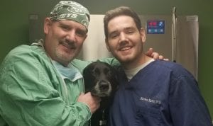
Dr. Kearns practices veterinary medicine from his Port Jefferson office and is pictured with his son Matthew and his dog Jasmine.

 February is National Pet Dental Health Month so I figured an article on dentistry is appropriate. A quick tooth anatomy reference: Everything below the gumline is considered the “root,” and everything above the gumline is considered the “crown.”

