Tags Posts tagged with "Arianna Maffei"

Arianna Maffei

Arianna Maffei in her lab viewing a slice of brain that shows the mark of the infusion in the gustatory cortex (identified by the red and yellow). This is one of the images which helped the researchers confirm their study data. Photo courtesy of Stony Brook University

Study findings reveal a specific signal in one brain region that may hold the key

Researchers at Stony Brook University used genetic manipulation in a laboratory brain model to demonstrate that neurosteroids, signals involved in mood regulation and stress, can reduce the sensitivity and preference for sweet tastes when elevated within the gustatory cortex – a region in the brain most involved with taste. Their findings are published in Current Biology.

According to senior author Arianna Maffei, PhD, Professor in the Department of Neurobiology and Behavior, studies in humans suggest that the preference for certain foods influences how much we eat and that decreased sensitivity to taste is often associated with overconsumption, which may lead to obesity. Currently there is limited knowledge of how brain activity contributes to the differences in taste preference.

Determining the relationship between brain activity, taste and eating habits is difficult in humans because available technology for measuring changes in brain activity does not have sufficient resolution to identify biological mechanisms. However, scientists can accurately monitor brain activity in lab mice while measuring their taste preferences.

As the biology of taste is very similar in all mammals, this approach can shed light on the human brain and taste.

In their murine model, the research team investigated neural circuits regulating the preference for sweet taste in adult brains. Their work focused on the effect of the neurosteroid allopregnanolone, which is known to be elevated in people affected by obesity.

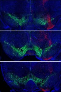
This neurosteroid modulates brain activity by increasing tonic inhibitory circuits mediated by a specific type of GABA receptor. The team demonstrated that these GABA receptors are present in excitatory and inhibitory neurons in the gustatory cortex.

They infused allopregnanolone locally into the gustatory cortex of the mice to activate neurosteroid-sensitive GABA receptors. This manipulation reduced the model’s sensitivity and preference for sweet taste. Then they used genetic tools to remove neurosteroid sensitive GABA receptors locally, only in the gustatory cortex. This manipulation eliminated the preference for sweet taste over water.

“This reduced sensitivity and preference for sweet taste was even more prominent if the receptors were selectively removed only from inhibitory gustatory cortex neurons. Indeed, in this case mice were practically unable to distinguish sugared water from water,” explains Maffei.

Their approach confirmed that a specific type of GABA receptor is the target of neurosteroid activity and is essential for fine-tuning sensitivity and preference for sweet taste.

Maffei says their findings illustrate the fascinating ways the mammalian brain contributes to the taste experience and reveals a specific signal in a specific brain region that is essential for sensitivity to sweet taste.

Ongoing research with the models is exploring whether neurosteroids only regulate sweet taste sensation or contribute to the perception of other tastes, and/or how changes in taste sensitivity influences eating.

The research was supported by several grants from the National Institute for Deafness and Communication Disorder (NIDCD) branch of the National Institutes of Health (NIH) and was supported by NIH grants R01DC019827, R01DC013770, R01DC015234, F31 DC019518 and UF1NS115779.

The authors are members of Stony Brook University’s College of Arts and Science (Yevoo and Maffei) and of the Renaissance School of Medicine (Fontanini).


Arianna Maffei, left, and lead author Hillary Schiff, in Maffei’s Neurobiology lab at Stony Brook. Photo by Josh F. Kogan

Findings from a Stony Brook University research team published in Science Advances

 Have you ever thought about how your food preferences came to be? Food preferences arise as a consequence of experience with food and shape eating habits and cultural identity, as Jean-Anthelme Brillat-Savarin nicely summarized in this quote from his work “The Physiology of Taste” published in 1825: “Tell me what you eat: I will tell you what you are.”

A new study by Stony Brook University researchers brought this concept to the scientific level and showed there is indeed a strong relationship between what we eat early in life, as babies or young children, and food preferences in adults. This relationship depends the effects of our early experience with food has on the brain. The research, published in Science Advances, highlights the importance of early exposure to a variety of tastes and identifies the neural basis regulating preferences for favorite foods, providing important new information about the relationship between nutrition and brain function.

Previous investigations of human infants hinted at the effect of early taste experience on food preference later in life. However, no previous study examined the neural bases of this phenomenon. This study looks at the neural bases of taste preference and provides findings that could form a basis to understanding the neural processes involved in taste preference.

The biology of the gustatory system is similar across all mammals. By using a murine model, the research team from the Department of Neurobiology and Behavior in the Renaissance School of Medicine exposed groups of mice to a variety of taste solutions for one week. They exposed groups as either weanlings (early exposure) or as adults (late exposure). After the one week experiencing a variety of tastes, they returned the groups to their regular diet, which is contains balanced nutrients but with is not rich in taste. For comparison, a control group of mice was raised only on the regular, blander diet.

“Our research is directed at assessing whether and how the gustatory experience and diet influence brain development. This study shows that the gustatory experience has fundamental effects on the brain. The next steps will be to determine how different diets such as a high fat, or a high sugar or high salt, may influence taste preferences and neural activity, ” explains Arianna Maffei, PhD, Senior Author and Professor in the Department of Neurobiology and Behavior.

Maffei, lead author Hillary Schiff, and colleagues increased taste variety in the healthy diets of mice and found that the development of neural circuits and taste preference are influenced by all aspects of the gustatory experience: sensations in the mouth, smell, and gut-brain relations.

Several weeks after exposing the groups to the one-week taste variety, the investigators measured preference for a sweet solution compared to water. Mice who experienced taste variety early in life had a stronger preference for sweet tastes in adulthood compared to the control group. This change preference depended on a combination of taste, smell, and gut-to-brain signals, and was specific to early exposure taste. Mice exposed to taste variety as adults did not show different sweet preferences from their age-matched control group. These results indicated that taste experience influences preference, but only if given within a restricted time window.

The researchers also recorded the activity of neurons in the gustatory cortex of all the subjects. This part of the brain is involved in taste perception and decisions about ingesting or rejecting foods. The recorded activity showed that the shift in sweet preference was associated with differences in the activity of inhibitory neurons of adult mice.

This led to the question of whether manipulating these inhibitory neurons in adulthood can re-open the window of sensitivity to the taste experience.

To answer this question the research team injected a substance into the gustatory cortex that breaks down perineuronal nets, which are webs of proteins that accumulate around inhibitory neurons early in life. Once established, these nets play a key role in limiting plasticity – the ability to change in response to stimuli at inhibitory synapses.

When adult mice without perineuronal nets in the gustatory cortex were exposed to the taste variety, they showed a similar change in sweet preference as the group exposed earlier in life. This manipulation “rejuvenated” inhibitory synapses in the gustatory cortex and restored plasticity in response to taste experience, which confirmed the importance of maturation and plasticity in inhibitory circuits for the development of taste preference in the experimental model.

“It was striking to discover how long-lasting the effects of early experience with taste were in the young groups,” says Schiff. “The presence of a ‘critical period’ of the life cycle for the development of taste preference was a unique and exciting discovery. The prevailing view from other studies prior to this finding was that taste does not have a defined window of heightened sensitivity to experience like other sensory systems such as vision, hearing, and touch.”

The authors maintain that while the study was done in mice, the results inform scientists on the fundamental biological aspects of experiences with taste that extends beyond animal models and to humans.

“The development of taste preference requires a full gustatory experience,” adds Maffei. “This includes the detection of taste in the mouth, its association with smell and the activation of gastrointestinal sensations. All these aspects influence the activity of brain circuits, promoting their healthy development.”

Regarding humans, Maffei points out that we often favor food from our childhood, highlighting important cultural aspects of our taste experience. Additionally, in the public health realm several neurodevelopmental and neurodegenerative disorders are often associated with hyper- or hyposensitivity to gustatory stimuli, suggesting links between taste and brain function in health and disease .

“Expanding our knowledge of the developmental neural circuits for tastes – as studies like this do – will contribute to our understanding of food choices, eating disorders, and diseases associated with brain disorders,” emphasizes Maffei.

Schiff, Maffei, and collaborators conclude that their overall experimental results establish a fundamental link between the gustatory experience, sweet preference, inhibitory plasticity, circuit function, and the importance of early life nutrition in setting taste preferences.

The research was supported by several grants from the National Institute on Deafness and Other Communication Disorders and from the National Institute of Neurological Disorders and Stroke at the National Institutes of Health.

Olivia Swanson
Arianna Maffei

The role of neuron and dopamine loss in Parkinson’s Disease (PD) has long been recognized by neuroscientists. However, how dopaminergic modulation affects brain regions involved in the control of voluntary movement remains a subject of investigation.

Researchers in the Department of Neurobiology and Behavior in the College of Arts and Sciences and the Renaissance School of Medicine at Stony Brook University, used an experimental model to demonstrate that a loss of midbrain dopaminergic centers impairs the ability of the primary motor cortex neurons to transform inputs into appropriate output. The finding, published in eNeuro, supports a new line of research regarding the origins of changes in the motor cortex and its role during PD.

Patients with PD show abnormal activity in the motor cortex, which to date remains difficult to explain. Scientists have proposed that motor cortex dysfunction in PD may come from loss of direct dopaminergic innervation of the cortex, or, alternatively, it could arise as a consequence of basal ganglia pathology.

Dopamine neurons are vital to a healthy brain, but they degenerate in Parkinson’s Disease. This coronal section of the ventral part of the brain visualizes midbrain dopamine neurons in a healthy brain. Green: dopamine neurons. Red: axons from the motor cortex. Blue: all neurons, cell bodies. Image from Olivia Swanson

“Our study shows that the changes in excitability of motor cortex neurons very likely are due to basal ganglia pathology and not loss of direct dopaminergic innervation of the motor cortex,” says Arianna Maffei, PhD, Professor of Neurobiology and Behavior. “The results we showed support the idea that changes in motor cortex activity due to loss of dopamine are very important for the pathophysiology of PD. This adds to our current knowledge and points to the motor cortex as a potential novel site for intervention.”

The research team assessed how the loss of dopamine affects the input/output function of neurons in the motor cortex. They tested three different ways to reduce dopamine signaling to ask how motor cortex dysfunction may arise: 1) Used pharmacology to block the receptors selectively in the motor cortex 2) Injected a toxin that kills dopaminergic neurons in the midbrain to induce basal ganglia pathology, and 3) Used the same toxin to eliminate dopamine neuron axons in the motor cortex to test the possibility that loss of dopaminergic input to the motor cortex may be responsible for its dysfunction.

Professor Maffei explains that the idea behind these approaches was to dissect out the circuit mechanisms underlying loss of function in the motor cortex and possibly use these data to better understand PD pathophysiology.

Overall, the research demonstrated that diminished dopamine signaling, whether acute or chronic, has profound effects on the excitability of primary motor cortex neurons.

The authors believe the results should spur additional research that focuses on the primary motor cortex as an additional site of intervention to treat motor symptoms and improve outcomes in PD patients.