Medical Compass

It is very important to take white-coat hypertension seriously. Stock photo
As many as 30 percent of patients experience this phenomenon

By David Dunaief, M.D.

Dr. David Dunaief

White-coat hypertension (high blood pressure) is defined as blood pressure that is elevated to at least 140/90 mm Hg at a physician’s office, but “normal” when measured at home. The blood pressure considered normal at home for most Americans is less than 135/85 mm Hg. This is a real phenomenon caused by the anxiety or stress of being in a doctor’s office. It is also known as “isolated office hypertension.”

About 15 to 30 percent of patients experience white-coat hypertension (1). However, when the diastolic (bottom number) blood pressure is greater than 105 mm Hg, it is unlikely to be simply caused by doctor’s office-related stress (2).

Consequences

What are the consequences of white-coat hypertension? The first challenge is that physicians may overtreat it, prescribing medications that lead to low blood pressure when not in the office. Alternately, we sometimes discount it because it seems benign or harmless. However, some studies show that it may increase the risk of sustained hypertension, which is a major contributor to developing cardiovascular disease — heart disease and stroke.

It is very important to take white-coat hypertension seriously because Centers for Disease Control and Prevention data show that the percentage of adults age 20 and over with hypertension reached 33.5 percent in the 2013-14 period (3).

What can be done?

What can be done about white-coat hypertension? Well, it does not need to be treated with medication, except potentially in elderly patients (over 80 years of age) but should involve lifestyle modifications, including dietary changes, stress reduction and exercise. In terms of diet, increased beet juice, green leafy vegetables and potassium, as well as decreased sodium intake may be important. You should monitor the blood pressure at home, taking multiple readings during the day, or by 24-hour ambulatory blood pressure readings, which require wearing a monitor. The latter provides the additional advantage of blood pressure readings during your sleep.

If you do monitor your blood pressure at home, the American Heart Association has suggestions on how to get the most accurate readings, such as measurements early in the morning before exercising and eating, as well as in the evening (4). You should also be comfortably seated, don’t cross your legs, and sit/relax for a few minutes before taking a reading. Let’s look at the evidence.

Risk of sustained high blood pressure

There were no substantial studies demonstrating any consequences from white-coat hypertension until 2005. Most previous studies on white-coat hypertension were not of long enough duration.

In the 2005 population-based Ohasama study, results showed that the participants who had white-coat hypertension were 2.9 times more likely to develop sustained hypertension, compared to those who had normal blood pressure in the doctor’s office (5). There were almost 800 participants involved in this study, with a mean age at the start of 56. What was really impressive about the study was its duration, with an eight-year follow-up. This gives a better sense of whether white-coat hypertension may develop into sustained hypertension. The researchers concluded that it may lead to a less than stellar outlook for cardiovascular prognosis.

Another study, published in 2009, reinforced these results. The PAMELA study showed that those with white-coat hypertension had about a 2.5-times increased risk of developing sustained high blood pressure, compared to those who had normal readings in all environments (6). There were 1,412 participants involved in the study, ranging in age from 25 to 74. Just like the previous study, an impressive aspect was the fact that there was a long follow-up period of 10 years. Thus, this was a substantial study, applicable to the general population over a significant duration.

Prevention of sustained hypertension

In a small, randomized controlled trial, beet juice was shown to reduce blood pressure significantly (7). Patients either were given 250 ml (about 8 ounces) of beet juice or comparable amounts of water. The patients who drank the beet juice saw an 11.2 mm Hg decrease in blood pressure, while those who drank water saw a 0.7 mm Hg reduction. This effect with the beet juice continued to remain significant. Even after 24 hours, there was a sustainable 7.2 mm Hg drop in blood pressure, compared to readings taken prior to drinking the juice. Although these results are encouraging, we need to study whether these effects can be sustained over the long term. Also, this study was done in patients with high blood pressure. I don’t know of any prevention studies done in patients with white-coat hypertension.

The researchers believe the effect is caused by high nitrate levels in beet juice that are converted to nitrite when it comes in contact with human saliva. Nitrite helps to vasodilate, or enlarge blood vessels, and thus helps to decrease blood pressure in a similar way as some antihypertensive (blood pressure) medications. The authors go on to surmise that green leafy vegetables offer protection from cardiovascular disease in part due to increased nitrite levels, similar to those in beet juice. 

A subsequent double-blind, placebo-controlled clinical trial with 68 hypertensive patients found that blood pressure was significantly reduced in the clinic and in home readings over a four-week period, when compared to nitrate-free beet juice (8).

If you have diabetes, prediabetes, a family history or a high risk for diabetes, I recommend eating beets instead, since drinking beet juice will raise your sugar levels.

Increasing potassium levels significantly through food sources, not supplements, has a profound effect in reducing blood pressure. In a study where 3,500 to 4,700 mg of potassium were consumed through foods, the systolic (top number) blood pressure was reduced by 7.1 mm Hg (9). We should be getting 4,700 mg of potassium daily, which equates to about 10 bananas daily. Almonds, raisins and green leafy vegetables, such as Swiss chard, also have significant amounts of potassium.

White-coat hypertension should not be neglected. It is important to monitor blood pressure at home for at least three days with multiple readings, and then send them to your physician for review. Though patients don’t need to be on blood pressure medications at this stage, it does not mean you should be passive about the process. Make lifestyle modifications to reduce your risk of developing sustained hypertension.

References:

(1) Hypertension. 2013;62:982-987, originally published Nov 13, 2013. (2) J Hypertens. 2001;19(6):1015. (3) cdc.gov. (4) Am Fam Physician. 2005 Oct 1;72(7):1391-1398. (5) Arch Intern Med. 2005 Jul 11;165(13):1541-1546. (6) Hypertension. 2009; 54: 226-232. (7) Hypertension. Online 2013; April 15. (8) Hypertension. 2015 Feb; 65(2): 320–327. (9) BMJ. 2013 Apr 3;346:f1378. 

Dr. Dunaief is a speaker, author and local lifestyle medicine physician focusing on the integration of medicine, nutrition, fitness and stress management. For further information, visit www.medicalcompassmd.com or consult your personal physician.

Lifestyle modifications play a role in reducing the risk of developing dementia. Stock photo
Managing biological age through diet can reduce risk

By David Dunaief, M.D.

Dr. David Dunaief

Dementia may be diagnosed when someone experiences loss of memory plus loss of another faculty, such as executive functioning (decision-making) or language abilities (speaking, writing or reading). The latter is known as aphasia. Alzheimer’s disease is responsible for approximately 60 to 80 percent of dementia cases (1).

Unfortunately, there are no definitive studies that show reversal or a cure for Alzheimer’s disease. This is why prevention is central to Alzheimer’s — and dementia in general.

In terms of dementia, there is good news and some disappointing news.

We will start with the good news. Though chronological age is a risk factor that cannot be changed, biological age may be adjustable. There are studies that suggest we may be able to prevent dementia through the use of both lifestyle modifications and medications.

Telomeres’ length and biological age

Biological age may be different from chronological age, depending on a host of environmental factors that include diet, exercise and smoking. There are substances called telomeres that are found at the ends of our chromosomes. They provide stability to this genetic material. As our telomeres get shorter and shorter, our cellular aging and, ultimately, biological aging, increases.

In a preliminary case-control study, dementia patients were shown to have significantly shorter telomere length than healthy patients (2). Interestingly, according to the authors, men have shorter telomere length and may be biologically older by four years than women of the same chronological age. The researchers caution that this is a preliminary finding and may not have clinical implications.

What I find most intriguing is that intensive lifestyle modifications increased telomere length in a small three-month study with patients who had low-risk prostate cancer (3). By adjusting their lifestyles, study participants were potentially able to decrease their biological ages.

Diet’s effect

Lifestyle modifications play a role in many chronic diseases and disorders. Dementia is no exception. In a prospective observational study, involving 3,790 participants, those who had the greatest compliance with a Mediterranean-type diet demonstrated a significant reduction in the risk of Alzheimer’s disease, compared to the least compliant (4). Participants were over the age of 65, demographics included substantial numbers of both black and white participants, and there was a mean follow-up of 7.6 years. Impressively, those who adhered more strictly to the diet performed cognitively as if they were three years younger, according to the authors.

Beta-carotene and vitamin C effect

In a small, preliminary case-control study (disease vs. healthy patients), higher blood levels of vitamin C and beta-carotene significantly reduced the risk of dementia, by 71  and 87 percent, respectively (5). The blood levels were dramatically different in those with the highest and lowest blood levels of vitamin C (74.4 vs. 28.9 µmol/L) and beta-carotene (0.8 vs. 0.2 µmol/L).

The reason for this effect may be that these nutrients help reduce oxidative stress and thus have neuroprotective effects, preventing the breakdown of neurons. This study was done in the elderly, average 78.9 years old, which is a plus, since as we age we’re more likely to be afflicted by dementia.

It is critically important to delineate the sources of vitamin C and beta-carotene in this study. These numbers came from food, not supplements. Why is this important? First, beta-carotene is part of a family of nutrients called carotenoids. There are at least 600 carotenoids in food, all of which may have benefits that are not achieved when taking beta-carotene supplements. Second of all, beta-carotene in supplement form may increase the risk of small-cell lung cancer in smokers (6).

Foods that contain beta-carotene include fruits and vegetables such as berries; green leafy vegetables; and orange, red or yellow vegetables like peppers, carrots and sweet potato. It may surprise you, but fish also contains carotenoids. In my practice, I test for beta-carotene and vitamin C as a way to measure nutrient levels and track patients’ progress when they are eating a nutrient-dense diet. Interestingly, many patients achieve more than three times higher than the highest beta-carotene blood levels seen in this small study.

Impact of high blood pressure medications

For those patients who have high blood pressure, it is important to know that not all blood pressure medications are created equal. When comparing blood pressure medications in an observational study, two classes of these medications stood out. Angiotensin II receptor blockers (known as ARBs) and angiotensin-converting enzyme inhibitors (known as ACE inhibitors) reduce the risk of dementia by 53 and 24 percent respectively, when used in combination with other blood pressure medications.

Interestingly, when ARBs were used alone, there was still a 47 percent reduction in risk; however, ACE inhibitors lost their prevention advantage. High blood pressure is a likely risk factor for dementia and can also be treated with lifestyle modifications (7). Otherwise, ARBs or ACE inhibitors may be the best choices for reducing dementia risk.

Ginkgo biloba disappoints

Ginkgo biloba, a common herbal supplement taken to help prevent dementia, may have no benefit. In the GuidAge study, ginkgo biloba was shown to be no more effective than placebo in preventing patients from progressing to Alzheimer’s disease (8). This randomized controlled trial, considered the gold standard of study designs, was done in elderly patients over a five-year period with almost 3,000 participants. There was no difference seen between the treatment and placebo groups. This reinforces the results of an earlier study, Ginkgo Evaluation of Memory trial (9). Longer studies may be warranted. The authors stressed the importance of preventive measures with dementia.

Fish oil: not the last word

Many of us take fish oil supplements in the hope of preventing dementia. However, in a meta-analysis (a group of three randomized controlled trials), the results did not show a difference between treatment groups and placebo in older patients taking fish oil with omega-3 fatty acids (10). The authors stress that this is not the final word since studies have been mixed. 

The longest of the three studies was 40 months, yet may not have been long enough to see a beneficial effect. Also participants in the meta-analysis did not necessarily have low omega-3 levels at the beginning of the studies. This doesn’t necessarily mean fish oil doesn’t work for dementia prevention, it is just discouraging, as the authors emphasize. Fish consumption, however, has shown an inverse association with Alzheimer’s and dementia overall (11).

There may be ways to prevent dementia from occurring, whether through lifestyle modifications or through the selection of medications, if they are necessary.

References:

(1) www.uptodate.com. (2) Arch Neurol. 2012 Jul 23:1-8. (3) Lancet Oncol. 2008;9(11):1048-1057. (4) Am J Clin Nutr. 2011;93:601-607. (5) J Alzheimers Dis. 2012;31:717-724. (6) Am. J. Epidemiol. 2009; 169(7):815-828. (7) Neurology. 2005;64(2):277. (8) Lancet Neurol. 2012;11(10):851-859. (9) JAMA. 2008;300(19):2253-2262. (10) Cochrane Summaries online June 13, 2012. (11) Neurology. 2007;69(20):1921.

Dr. Dunaief is a speaker, author and local lifestyle medicine physician focusing on the integration of medicine, nutrition, fitness and stress management. For further information, visit www.medicalcompassmd.com or consult your personal physician.

Accumulating evidence supports an association between depression and inflammation. Stock photo
C-reactive protein is an important biomarker

By David Dunaief, M.D.

Dr. David Dunaief

Many of us have inflammation in our bodies, inflammation that is a potential underlying cause for a great number of diseases. Can we demonstrate the level of inflammation by measuring it? The answer is yes.

One of the most widely studied biomarkers for inflammation is high-sensitivity C-reactive protein (hsCRP), also referred to as CRP. High sensitivity means that we can measure levels as low as 0.3 mg/L more accurately.

What is the significance of the different levels? In heart disease, individuals who have levels lower than 1.0 mg/L are in the optimal range for low risk of inflammation. Levels of 1 to 3 mg/L represent the average risk range, and greater than 3.0 mg/L is a higher risk profile. Above 10.0 mg/L is less specific to heart disease, although still related, but more likely associated with other causes, such as infection and autoimmune diseases (1, 2). This biomarker is derived from the liver.

CRP is not specific to heart disease, nor is it definitive for risk of the disease. However, the upside is that it may be helpful with risk stratification, which helps us understand where we sit on a heart disease risk spectrum and with progression in other diseases, such as age-related macular degeneration, diabetic retinopathy, depression and autoimmune diseases. Let’s look at the evidence.

Age-related macular degeneration

Age-related macular degeneration (AMD) is the leading cause of blindness in patients over the age of 65 (3). Therefore, it is very important to help define risk stratification for this disease. In a prospective study, results showed that hsCRP levels were inversely associated with the risk of developing AMD. The group with an hsCRP greater than 3.0 mg/L had a 50 percent increased risk of developing overall AMD compared to the optimal group with hsCRP lower than 1.0 mg/L. But even more interestingly, the risk of developing neovascular, or wet, AMD increased to 89 percent in this high-risk group.

The significance of wet AMD is that it is one type of advanced-stage AMD that results in blindness. This study involved five studies where the researchers thawed baseline blood samples from middle-aged participants who had hsCRP levels measured. There were more than 2,000 participants with a follow-up as long as 20 years. According to the study’s authors, annual eye exams and lifestyle modifications, including supplements, may be able to stem this risk by reducing hsCRP.

These results reinforce those of a previous prospective study that showed that elevated hsCRP increased the risk of AMD threefold (4). This study utilized data from the Women’s Health Study, which involved over 27,000 participants. Like the study mentioned above, this one also defrosted blood samples from baseline and looked at follow-up incidence of developing AMD in initially healthy women.

The highest group had hsCRP levels over 5.2 mg/L. Additionally, when analyzing   similar cutoffs for high- and low-level hsCRP, as the above trial used, those with hsCRP over 3.0 had an 82 percent increased risk of AMD compared to those with an hsCRP of lower than 1.0 mg/L.

Diabetic retinopathy — a complication of diabetes

We know that diabetes affects approximately 10 percent of the U.S. population and is continuing to rise at a rapid rate. One of the complications of diabetes affects the retina (back of the eye) and is called diabetic retinopathy. This is a leading cause of vision loss (5). One of the reasons for the vision loss is macular edema, or swelling, usually due to rupture of tiny blood vessels below the macula, a portion of the back of the eye responsible for central vision.

The Diabetes Control and Complications Trial (DCCT), a prospective study involving over 1,400 Type 1 diabetes patients, showed an 83 percent increased risk of developing clinically significant macular edema in the group with the highest hsCRP levels compared to those with the lowest (6). Although these results were with Type 1 diabetes, patients with Type 2 diabetes are at equal risk of diabetic retinopathy if glucose levels, or sugars, are not well controlled.

Depression

Depression is a very difficult disease to control and is a tremendous cause of disability. If we can minimize the risk of complications and hospitalizations, this is probably the most effective approach.

Well, it turns out that inflammation is associated with depression. Specifically, in a prospective observational trial, rising levels of CRP had a linear relationship with increased risk of hospitalization due to psychological distress and depression (7).

In other words, compared to levels of less than 1 mg/L, those who were 1 to 3 mg/L, 3 to 10 mg/L and greater than 10 mg/L had increased risk from 30 to 84 to 127 percent, respectively. This study involved over 70,000 patients.

What can be done to reduce inflammation?

This is the key question, since we now know that hsCRP is associated with systemic inflammation. In the Nurses’ Health Study, a very large, prospective observational study, the Dietary Approaches to Stop Hypertension (DASH) diet decreased the risk of both heart disease and stroke, which is impressive. The DASH diet also decreases the levels of hsCRP significantly, which was associated with a decrease in clinically meaningful end  points of stroke and heart disease (8).

The DASH diet is nutrient dense with an emphasis on fruits, vegetables, nuts, seeds, legumes and whole grains and a de-emphasis on processed foods, red meats, sodium and sweet beverages.

Conclusion

As the evidence shows with multiple diseases, hsCRP is a very valuable nonspecific biomarker for inflammation in the body.

To stem the effects of inflammation, reducing hsCRP through lifestyle modifications and drug therapy may be a productive way of reducing risk, slowing progression and even potentially reversing some disease processes.

The DASH diet is a very powerful approach to achieving optimal levels of hsCRP without incurring potential side effects. This is a call to arms to have your levels measured, especially if you are at high risk or have chronic diseases such as heart disease, diabetes, depression and autoimmune diseases. HsCRP is a simple blood test with easy-to-obtain results.

References:

(1) uptodate.com. (2) Diabetes Technol Ther. 2006;8(1):28-36. (3) Prog Retin Eye Res. 2007 Nov;26(6):649-673. (4) Arch Ophthalmol. 2007;125(3):300-305. (5) Am J Ophthalmol. 2003;136(1):122-135. (6) JAMA Ophthalmol. 2013 Feb 7;131:1-8. (7) JAMA Psychiatry. 2013;70(2):176-184. (8) Arch Intern Med. 2008;168(7):713-720.

Dr. Dunaief is a speaker, author and local lifestyle medicine physician focusing on the integration of medicine, nutrition, fitness and stress management. For further information, visit www.medicalcompassmd.com or consult your personal physician.

Pedometers can be the first step to helping those with mild COPD. Stock photo
Lifestyle changes can reduce COPD exacerbations

By David Dunaief, M.D.

Dr. David Dunaief

COPD, or chronic obstructive pulmonary disease, is the third leading cause of mortality in the United States (1), although it’s not highlighted much in the layman’s press.

COPD is an umbrella term that includes emphysema, chronic bronchitis of more than three months for two consecutive years and/or chronic obstructive asthma. It is an obstructive lung disease that limits airflow. The three most common symptoms of the disease involve shortness of breath, especially on exertion, production of sputum and cough. This disease affects 6.7 percent of the U.S. population (2).

It tends to be progressive, meaning more frequent and severe exacerbations over time. Since it is a devastating and debilitating chronic disease with no cure, anything that can identify and prevent COPD exacerbations, as well as comorbidities (associated diseases), is critically important.

What are the traditional ways to reduce the risk of and treat COPD exacerbations? The most important step is to stop smoking, since 80 percent of COPD is related to smoking. Supplemental oxygen therapy and medications, such as corticosteroids, bronchodilators (beta-adrenergic agonists and anticholinergics) and antibiotics help to alleviate symptoms (3).

One of the underlying components of COPD may be chronic inflammation (4). Therefore, reducing inflammation may help to stem COPD exacerbations. There are several inflammatory biomarkers that could potentially help predict exacerbations and mortality associated with this disease, such as interleukin-6 (IL-6), C-reactive protein (CRP), leukocyte (white blood cell) count and fibrinogen (a clotting factor of the blood).

How do we reduce inflammation, which may contribute to exacerbations of this disease? Some drugs, such as statins, work partially by reducing inflammation. They may have a role in COPD. Lifestyle changes that include a high-nutrient, anti-inflammatory diet and exercise may also be beneficial. Let’s look at the evidence.

Biomarkers for inflammation

In a recent population-based study with over 60,000 participants, results show that as three biomarkers (CRP, leukocyte count and fibrinogen) were elevated, the risk of COPD exacerbation increased in a linear manner (5). In other words, the risk of frequent exacerbation increased 20, 70 and 270 percent within the first year as the number of elevated biomarkers increased from one to three, compared to patients who did not have biomarker elevations.

As time progressed beyond the first year of follow-up, risk exacerbation continued to stay high. Patients with all three biomarkers elevated for longer periods had a 150 percent increased risk of frequent exacerbations. These predictions were applicable to patients with stable and with mild COPD.

In an observational study, results showed that when the biomarker IL-6 was elevated at the start of the trial in stable COPD patients, the risk of mortality increased almost 2.7-fold (6). Also, after three years, IL-6 increased significantly. Elevated IL-6 was associated with a worsening of six-minute walking distance, a parameter tied to poor physical performance in COPD patients. However, unlike the previous study, CRP did not show correlation with increased COPD exacerbation risk. This was a small trial, only involving 53 patients. Therefore, the results are preliminary.

These biomarker trials are exciting for their potential to shape treatments based on level of exacerbation risk and mortality, creating more individualized therapies. Their results need to be confirmed in a randomized controlled trial (RCT). Many of these biomarkers mentioned in the two trials are identifiable with simple blood tests at major labs.

Statin effect

Statins have been maligned for their side effects, but their efficacy has been their strong suit. An observational trial showed that statins led to at least a 30 percent reduction in the risk of COPD exacerbations, with the effect based on a dose-dependent curve (7). In other words, as the dose increased, so did the benefit.

Interestingly, even those who had taken the statin previously saw a significant reduction in COPD exacerbation risk. The duration of statin use was not important; a short use of statins, whether presently or previously, had substantial benefit. 

However, the greatest benefit was seen in those who had been on a medium to high dose or were on the drug currently. The researchers believe that the mechanism of action for statins in this setting has to do with their anti-inflammatory and immune-modulating effects. This was a retrospective (backward-looking) study with over 14,000 participants. We will need a prospective (forward-looking) study and an RCT to confirm the results.

Exercise

Pedometers can be the first step to helping those with mild COPD. Stock photo

Exercise is beneficial for almost every circumstance, and COPD is no exception. But did you know that a pedometer might improve results? In a three-month study, those with mild COPD were much more successful at achieving exercise goals and reducing exacerbations and symptoms when they used pedometers, compared to the group given advice alone (8). Pedometers gave patients objective feedback on their level of physical activity, which helped motivate them to achieve the goal of walking 9,000 steps daily. This is a relatively easy way to achieve exercise goals and reduce the risk of COPD exacerbations.

When exercising, we are told to vary our exercise routines on a regular basis. One study demonstrates that this may be especially important for COPD patients (9). Results show that nonlinear periodization exercise (NLPE) training is better than traditional routines of endurance and resistance training in severe COPD patients. The goal of NLPE is to alter the time spent working out, the number of sets, the number of repetitions and the intensity of the workout on a regular basis.

This study was randomized, involved 110 patients and was three months in duration. Significantly more severe COPD patients achieved their exercise goals using NLPE rather than the traditional approach. The group that used NLPE also had an improved quality of life response. The researchers believe that compliance with an NLPE-type program is mostly likely going to be greater because patients seem to enjoy it more.

Chronic inflammation may play a central role in COPD exacerbation. Nonspecific inflammatory biomarkers are potentially valuable for providing a more personalized approach to therapy. Drugs that can control inflammation, such as statins, show promise. But don’t forget the importance of lifestyle changes, such as quitting smoking and committing to an exercise regimen that is varied and/or involves the use of a pedometer. And potentially a high-nutrient, anti-inflammatory diet will also contribute positively to reducing the frequency and severity of COPD exacerbations.

References:

(1) Natl Vital Stat Rep. 2011 Dec.;59(10):1-126. (2) cdc.gov. (3) N Engl J Med. 2002;346:988-994. (4) www.goldcopd.org. (5) JAMA. 2013;309:2353-2361. (6) Respiratory Research. 2013;14:24. (7) Am J Med. 2013 Jul;126:598-606. (8) ATS 2013 International Conference: Abstract A1360. (9) Am J Respir Crit Care Med. 2013; online Feb. 28.

Dr. Dunaief is a speaker, author and local lifestyle medicine physician focusing on the integration of medicine, nutrition, fitness and stress management. For further information, visit www.medicalcompassmd.com or consult your personal physician.

In addition to bananas, plenty of other foods are high in potassium. Above are just a few examples. Stock photo
Most Americans don’t consume enough potassium

By David Dunaief M.D.

Dr. David Dunaief

One of the most popular food additives is also one of the most dangerous: salt. We need salt, but not in excess. On the other hand, potassium is beneficial in our diet. However, we have the opposite problem with potassium: It is under-consumed.

More than 90 percent of people consume far too much sodium, with salt being the primary culprit (1). Sodium is found in foods that don’t even taste salty. Bread and rolls are the primary offenders, since we eat so much of them. Other foods with substantial amounts of sodium are cold cuts and cured meats, cheeses, pizza (which has both bread and cheese), fresh and processed poultry, soups, meat dishes, pastas and snack foods. Foods that are processed and those prepared by restaurants are where most of our consumption occurs (2).

By contrast, only about 2 percent of people get enough potassium from their diets (3). According to one study, we would need to consume about eight sweet potatoes or 10 bananas each day to reach appropriate levels. Why is it important to reduce sodium and increase potassium? A high sodium-to-potassium ratio increases the risk of cardiovascular disease by 46 percent, according to the study, which looked at more than 12,000 Americans over almost 15 years (4). In addition, both may have significant impacts on blood pressure and cardiovascular disease.

To improve our overall health, we need to tip the sodium-to-potassium scales, consuming less sodium and more potassium. Let’s look at the evidence.

Reduced sodium

There are two studies that illustrate the benefits of reducing sodium in high blood pressure and normotensive (normal blood pressure) patients, ultimately preventing cardiovascular disease, including heart disease and stroke.

The first study used the prestigious Cochrane review to demonstrate that blood pressure is reduced by a significant mean of −4.18 mm Hg systolic (top number) and −2.06 mm Hg diastolic (bottom number) involving both normotensive and hypertensive participants (5). When looking solely at hypertensive patients, the reduction was even greater, with a systolic blood pressure reduction of −5.39 mm Hg and a diastolic blood pressure reduction of −2.82 mm Hg.

This was a meta-analysis (a group of studies) that evaluated data from randomized clinical trials, the gold standard of studies. There were 34 trials reviewed with more than 3,200 participants. Salt was reduced from 9 to 12 grams per day to 5 to 6 grams per day. These levels were determined using 24-hour urine tests. The researchers believe there is a direct linear effect with salt reduction. In other words, the more we reduce the salt intake, the greater the effect of reducing blood pressure. The authors concluded that these effects on blood pressure will most likely result in a decrease in cardiovascular disease.

In the second study, a meta-analysis of 42 clinical trials, there was a similarly significant reduction in both systolic and diastolic blood pressures (6). This meta-analysis included adults and children. Both demographics saw a reduction in blood pressure, though the effect, not surprisingly, was greater in adults. Interestingly, an increase in sodium caused a 24 percent increased risk of stroke incidence but, more importantly, a 63 percent increased risk of stroke mortality. The risk of mortality from heart disease was increased as well, by 32 percent.

In an epidemiology modeling study, the researchers projected that either a gradual or instantaneous reduction in sodium would save lives (7). For instance, a modest 40 percent reduction over 10 years in sodium consumed could prevent 280,000 premature deaths. These are only projections, but in combination with the above studies may be telling. The bottom line is: decrease sodium intake by almost half and increase potassium intake from foods.

Potassium’s positive effects

When we think of blood pressure, sodium comes to mind, but not enough attention is given to potassium. The typical American diet doesn’t contain enough of this mineral.

In a meta-analysis involving 32 studies, results showed that as the amount of potassium was increased, systolic blood pressure decreased significantly (8). When foods containing 3.5 to 4.7 grams of potassium were consumed, there was an impressive −7.16 mm Hg reduction in systolic blood pressure with high blood pressure patients. Anything more than this amount of potassium did not have any additional benefit. Increased potassium intake also reduced the risk of stroke by 24 percent. This effect was important. If this does not sound like a large reduction, consider that, by comparison, aspirin has been shown to reduce the risk of stroke by 20 percent.

The reduction in blood pressure was greater with increased potassium consumption than with sodium restriction, although there was no head-to-head comparison done. The good news is that potassium is easily attainable in the diet. Foods that are potassium rich include bananas, sweet potatoes, almonds, raisins and green leafy vegetables such as Swiss chard.

Lowering sodium intake may have far-reaching benefits, and it is certainly achievable. We need to reduce our intake and give ourselves a brief period to adapt — it takes about six weeks to retrain our taste buds, once we reduce our sodium intake. We can also improve our odds by increasing our dietary potassium intake, which also has a substantial beneficial effect, striking a better sodium-to-potassium balance.

References:

(1) Am J Clin Nutr. 2012 Sep;96(3):647-657. (2) www.cdc.gov. (3) Am J Clin Nutr. 2012 Sep;96(3):647-657. (4) Arch Intern Med. 2011;171(13):1183-1191. (5) BMJ. 2013 Apr 3;346:f1325. (6) BMJ. 2013 Apr 3;346:f1326. (7) Hypertension. 2013; 61: 564-570. (8) BMJ. 2013; 346:f1378.

Dr. Dunaief is a speaker, author and local lifestyle medicine physician focusing on the integration of medicine, nutrition, fitness and stress management. For further information, visit www.medicalcompassmd.com or consult your personal physician.

Concentrate on lifestyle modifications like going for walks if you want to see potentially disease-modifying effects. Stock photo
Diet and exercise changes may slow progression

By David Dunaief, M.D.

Not surprisingly, osteoarthritis is widespread. The more common joints affected are the knees, hips and hands. There are three types of treatment for this disease: surgery, involving joint replacements of the hips or knees; medications; and nonpharmacologic approaches. The most commonly used first-line medications are acetaminophen and nonsteroidal anti-inflammatory drugs, such as ibuprofen. Unfortunately, medications mostly treat the symptoms of pain and inflammation.

However, the primary objectives in treating osteoarthritis should also include improving quality of life, slowing progression of the disease process and reducing its disabling effects (1).

Dairy and milk

When we think of dairy, specifically milk, there are two distinct camps: One believes in the benefits, and the other thinks it may contribute to the disease. In this case they both may be at least partly correct. In the Osteoarthritis Initiative study, an observational study of over 2,100 patients, results showed that low-fat (1 percent) and nonfat milk may slow the progression of osteoarthritis (2). The researchers looked specifically at joint space narrowing that occurs in those with affected knee joints. Radiographic imaging changes were used at baseline and then to follow the patients for up to 12 to 48 months for changes. Compared to those who did not drink milk, patients who did saw significantly less narrowing of knee joint space.

Was it a dose-dependent response? Not necessarily. Specifically, those who drank less than three glasses/week and those who drank four to six glasses/week both saw slower progression of joint space narrowing of 0.09 mm. Seven to 10 glasses/week resulted in a 0.12 mm preservation. However, those who drank more than 10 glasses/week saw less beneficial effect, 0.06 mm preservation compared to those who did not drink milk. Interestingly, there was no benefit seen in men or with the consumption of cheese or yogurt.

However, there are significant flaws with this study. First, the patients were only asked about their dietary intake of milk at baseline; therefore their consumption could have changed during the study. Second, there was a recall bias; patients were asked to recall their weekly milk consumption for the previous 12 months before the study began. I don’t know about you, but I can’t recall my intake of specific foods for the last week, let alone for the past year. Third, there could have been confounding factors, such as orange consumption.

Oddly, this was not a dose-response curve, since the most milk consumption had less beneficial effect than lower amounts. Also, why were these effects only seen in women? Finally, researchers could not explain why low-fat or nonfat milk had this potential benefit, but cheese was detrimental and yogurt did not show benefit. We are left with more questions than answers.

Would I recommend consuming low-fat or nonfat milk? Not necessarily, but I may not dissuade osteoarthritis patients from drinking it. There are very few approaches that slow the progression of joint space narrowing.

Vitamin D

Over the last five years or so, the medical community has gone from believing that vitamin D was potentially the solution to many diseases to wondering whether, in some cases, low levels were indicative of disease, but repletion was not a change-maker. Well, in a randomized controlled trial (RCT), the gold standard of studies, vitamin D had no beneficial symptom relief nor any disease-modifying effects (3). This two-year study of almost 150 men and women raised blood levels of vitamin D on average to 36 ng/ml, which is considered respectable. Researchers used MRI and X-rays to track their results.

Glucosamine

There is raging debate about whether glucosamine is an effective treatment for osteoarthritis. In the latest installment, there was an RCT, the results of which showed that glucosamine hydrochloride was not effective in treating osteoarthritis (4). In the trial, 201 patients with either mild or moderate knee pain drank diet lemonade with or without 1,500 mg of glucosamine hydrochloride.

There was no difference in cartilage changes in the knee nor in pain relief in those in the placebo or treatment groups over a six-month duration. Bone marrow lesions also did not improve with the glucosamine group. The researchers used 3T MRI scans (an advanced radiologic imaging technique) to follow the patients’ disease progression. This does not mean that glucosamine does not work for some patients. Different formulations, such as glucosamine sulfate, were not used in this study.

Weight

This could not be an article on osteoarthritis if I did not talk about weight. Do you remember analogies from the SATs? Well here is one for you: Weight loss, weight loss, weight loss is to osteoarthritis as location, location, location is to real estate. In a study involving 112 obese patients, there was not only a reduction of knee symptoms in those who lost weight, but there was also disease modification, with reduction in the loss of cartilage volume around the medial tibia (5).

On the other hand, those who gained weight saw the inverse effect. A reduction of tibial cartilage is potentially associated with the need for knee replacement. The relationship was almost one to one; for every 1 percent of weight lost, there was a 1.2 mm³ preservation of medial tibial cartilage volume, while the exact opposite was true with weight gain.

Exercise and diet

In a study, diet and exercise trumped the effects of diet or exercise alone (6). Patients with osteoarthritis of the knee who lost at least 10 percent of their body weight experienced significant improvements in function and a 50 percent reduction in pain, as well as reduction in inflammation, compared to those who lost 5 to 10 percent and those who lost less than 5 percent. This study was a well-designed, randomized controlled single-blinded study with a duration of 18 months.

Researchers used a biomarker — IL6 — to measure inflammation. The diet and exercise group and the diet-only group lost significantly more weight than the exercise-only group, 23.3 and 19.6 pounds versus 4 pounds. The diet portion consisted of a meal replacement shake for breakfast and lunch and then a vegetable-rich, low-fat dinner. Low-calorie meals replaced the shakes after six months. The exercise regimen included one hour of a combination of weight training and walking with alacrity three times per week.

Therefore, concentrate on lifestyle modifications if you want to see potentially disease-modifying effects. These include both exercise and diet. In terms of low-fat or nonfat milk, while the study had numerous flaws, if you drink milk, you might continue for the sake of osteoarthritis, but stay on the low end of consumption. And remember, the best potential effects shown are with weight loss and with a vegetable-rich diet.

References:

(1) uptodate.com. (2) Arthritis Care Res online. 2014 April 6. (3) JAMA. 2013;309:155-162. (4) Arthritis Rheum online. 2014 March 10. (5) Ann Rheum Dis online. 2014 Feb. 11. (6) JAMA. 2013;310:1263-1273.

Dr. Dunaief is a speaker, author and local lifestyle medicine physician focusing on the integration of medicine, nutrition, fitness and stress management. For further information, visit www.medicalcompassmd.com or consult your personal physician.
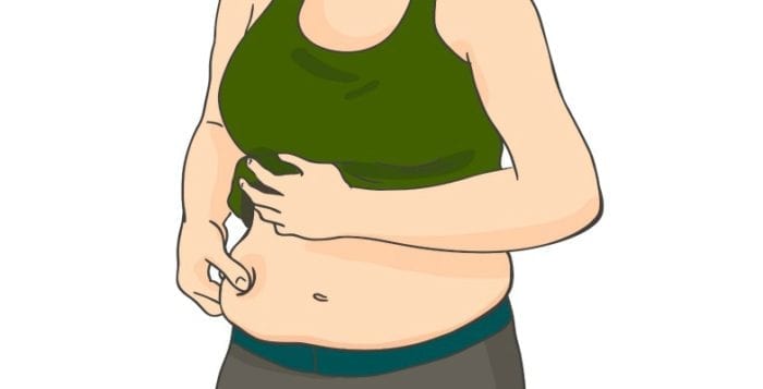
Season allergies are triggered by pollen from trees, grass and weeds. Stock photo

By David Dunaief, M.D.

Dr. David Dunaief

After winter ends, we look forward to mild temperatures. The days get longer, trees and flowers bud and bloom, and grass becomes lush and green. It seems like heaven. But for people who suffer from seasonal allergic rhinitis, hay fever, seasonal allergies or whatever you would like to call it, life can be downright miserable. You probably can rate an allergy season with your own built-in personal barometer, the sneeze factor. How many times are you, your friends or your colleagues sneezing?

Approximately 20 million U.S. adults have had a diagnosis of seasonal allergies within the past year, just a little over 8 percent of the population, and an additional 6.1 million children have this disorder, or about 8.4 percent, according to the Centers for Disease Control and Prevention (1). Sadly, considering the number of people it affects, only a paltry amount of research has been published.

The triggers for seasonal allergies are diverse. They include pollen from leafy trees and shrubs, lush grass and beautiful flowering plants, as well as weeds, with the majority from ragweed (mostly in the fall) and fungus (summer and fall) (2).

What sparks allergies within the body? A chain reaction occurs in seasonal allergy sufferers. When foreign substances such as allergens (pollen, in this case) interact with immunoglobulin E (IgE), antibodies that are part of our immune system, it causes mast cells in the body’s tissues to degrade and release inflammatory mediators. These include histamines, leukotrienes and eosinophils in those who are susceptible. In other words, it is an allergic inflammatory response.

The revved up immune system then responds with sneezing; red, itchy and watery eyes; scratchy throat; congestion; sinus headaches; postnasal drip; runny nose; diminished taste and smell; and even coughing (3). Basically, it emulates a cold, but without the virus. If symptoms last more than 10 days and are recurrent, then it is more than likely you have allergies.

Risk factors for seasonal allergies are tied most strongly to family history and to having other personal allergies, such as eczema or food allergies, but also may include cigarette exposure, being male and, possibly, diet (4). If allergic rhinitis is not properly treated, complications such as ear infections, sinusitis, irritated throat, insomnia, chronic fatigue, headaches and even asthma can result (5).

To treat allergic rhinitis, we have a host of medications from classes including intranasal glucocorticoids (steroids), oral antihistamines, allergy shots, decongestants, antihistamine and decongestant eye drops and leukotriene modifiers (second-line only).

The best way to treat allergy attacks is to prevent them, but this is an arduous process that can mean closing yourself out from the enjoyment of spring by literally closing the windows, using the air-conditioning, and using recycling vents in your car.

The guidelines for treating seasonal allergic rhinitis with medications suggest that intranasal corticosteroids (steroids) should be used when quality of life is affected. If there is itchiness and sneezing, then second-generation oral antihistamines may be appropriate (6). Two well-known inhaled steroids that do not require a prescription are Nasocort (triamcinolone) and Flonase (fluticasone propionate). There does not seem to be a significant difference between them (7). While inhaled steroids are probably most effective in treating and preventing symptoms, they need to be used every day and are not without side effects.

Oral antihistamines, on the other hand, can be taken on an as-needed basis. Second-generation antihistamines include loratadine (Claritin), cetirizine (Zyrtec) and fexofenadine (Allegra), and they have less sleepiness as a side effect than first-generation antihistamines.

Surprisingly good news

Seasonal allergic rhinitis may actually be beneficial for longevity. In a study involving more than 200,000 participants, results showed that those who had allergies had a 25 percent reduction in the risk of heart attacks, a 19 percent reduction in strokes, and a whopping 49 percent reduction in mortality (8). Remember two things: this is an observational trial, which means that it is not the best of trials, and don’t wish allergies on yourself. This effect may be at least partially attributable to the type of white blood cell expressed in the immune system.

In other words, type 2 T helper (Th2) lymphocytes (white blood cells) are elevated with allergies instead of type 1 T helper (Th1) lymphocytes. Why is this important? Th2 is known to decrease cardiovascular disease, while Th1 is known to possibly increase cardiovascular disease. Unfortunately, the same cannot be said about asthma, where cardiovascular events are increased by 36 percent.

Alternative treatments

Butterbur (Petasites hybridus), an herb, may not be just for migraines. There are several small studies that indicate its efficacy in treating hay fever. In fact, in one study, results showed that butterbur was as effective as cetirizine (Zyrtec) in treating this disorder (9). This was a small, randomized, controlled trial involving 131 patients.

In another randomized, controlled trial, results showed that high doses of butterbur — 1 tablet given three times a day — was significantly more effective than placebo (10). The side effects were similar in the placebo group and the butterbur group. The researchers used butterbur Ze339 (carbon dioxide extract from the leaves of Petasites hybridus L., 8 mg petasines per tablet) in the trial. The authors concluded that butterbur would be potentially useful for intermittent allergic rhinitis. The duration of treatment for this study was two weeks.

Still another study, this one a post-marketing study done as a follow-up to the previous study, showed that with butterbur Ze339, symptoms improved in 90 percent of patients with allergic rhinitis (11). Interestingly, anti-allergic medications were co-administered in about half of the patient population, with no additional benefit over butterbur alone. There were 580 patients in this study, and the duration was two weeks. Gastrointestinal upset occurred as the most common side effect in 3.8 percent of the population.

The caveats to the use of butterbur are several. First, the studies were short in duration. Second, the leaf extract used in these studies was free of pyrrolizidine alkaloids (PAs); this is very important since PAs may not be safe. Third, the dose was well-measured, which may not be the case with over-the-counter extracts. Fourth, you need to ask about interactions with your prescription medications.

Diet

While there are no significant studies on diet, there is one review of literature that suggests that a plant-based diet may reduce symptoms of allergies, specifically rhinoconjunctivitis, affecting the nose and eyes, as well as eczema and asthma. This is according to the International Study of Asthma and Allergies in Childhood study in 13- to 14-year-old teens (12). In my clinical practice, I have seen patients who suffer from seasonal allergies improve and even reverse the course of allergies over time with a vegetable-rich, plant-based diet, possibly due to an anti-inflammatory effect.

While allergies can be miserable, there are a significant number of over-the-counter and prescription options to help to reduce symptoms. Diet may play a role in the disease process by reducing inflammation, though there are no formal studies. There does seem to be promise with some herbs, especially butterbur. However, alternative supplements and herbs lack large, randomized clinical trials with long durations. Always consult your doctor before starting any supplements, herbs or over-the-counter medications.

References: (1) CDC.gov. (2) acaai.org/allergies/types/pollen-allergy. (3) Allergy Clin Immunol. 2003;112(6):1021-1031. (4) umm.edu. (5) J Allergy Clin Immunol. 2010;125(1):16-29. (6) Otolaryngol Head Neck Surg. online February 2, 2015. (7) Otolaryngol Head Neck Surg. 2003;129(1):16. (8) AAAAI 2014: Abstract 811. (9) BMJ 2002;324:144. (10) Arch Otolaryngol Head Neck Surg. 2004;130(12):1381-1386. (11) Adv Ther. 2006;23(2):373-384. (12) Eur Respir J. 2001;17(3):436-443.

Dr. Dunaief is a speaker, author and local lifestyle medicine physician focusing on the integration of medicine, nutrition, fitness and stress management. For further information, visit www.medicalcompassmd.com or consult your personal physician.

Symptoms of gallstones include dull or crampy abdominal pain that is exacerbated by meals and lasts one to five hours. Stock photo

By David Dunaief, M.D.

Dr. David Dunaief

Gallstones are a very common gastrointestinal disease; they affect up to 20 million Americans between the ages of 20 and 74, with a more than two-times increased occurrence in women than in men, according to the NHANES III survey (1). There are two types of gallstones, 80 percent of which are cholesterol stones and 20 percent of which are pigment stones.

Common symptoms

Gallstones may be asymptomatic; however, when gallstones block either the cystic or common bile ducts, symptoms occur. Symptoms include dull or crampy abdominal pain that is exacerbated by meals and lasts one to five hours. Jaundice, which includes yellowing of skin and eyes, is another symptom. Others include nausea and vomiting, rapid heart rate, hypotension (low blood pressure) and fever (2).

Tests used for diagnosis

Blood tests include complete blood count, where there may be a rise in white blood cells; liver enzymes; and the pancreatic enzymes lipase and amylase. In general, diagnostic tests that have more accuracy are the endoscopic ultrasonography (EUS) and endoscopic retrograde cholangiopancreatography (ERCP). However, these are invasive tests. Less accurate but noninvasive tests include abdominal X-ray, ultrasound and CAT scan (CT). The tests used also depend on where the stone may be located. Hepatobiliary (HIDA) scans are accurate if the stone is located in the cystic duct. And magnetic resonance retrograde cholangiopancreatography (MRCP) is used if the stone is thought to be located in the common bile duct (2).

What are the risk factors?

There are a multitude of risk factors. Some of these are modifiable, some others are not. The modifiable ones include obesity, measured by body mass index (BMI); rapid weight loss; fat consumption; hormone replacement therapy (HRT); oral contraceptives; decreased physical activity; Crohn’s disease; and certain drugs. One nonmodifiable risk factor is age; the older we get, the higher the risk, with age 40 being the demarcation line (3). Other risk factors are gender, with females being more predisposed; pregnancy; and family history (4). Let’s look at the evidence.

Obesity risks

Obesity may play an important role. Obesity is not age discriminant; it can impact both adults and children. The reason obesity is implicated is potentially due to bile becoming supersaturated (5). Bile is a substance produced in the liver and stored in the gallbladder. Bile aids in the digestion or breakdown of fats in the small intestines. Crystals may form, creating cholesterol gallstones from the bile.

Body mass index

A body mass index of greater than 30 kg/m² is considered obese. In a meta-analysis of two prospective, forward-looking observational trials, Copenhagen General Population Study and the Copenhagen City Heart Study, those in the highest quintile of BMI were almost three times as likely to experience symptomatic gallstones compared to those who were in the lowest quintile (6). The highest quintile was those who had a mean BMI of 32.5 kg/m² and thus were obese, whereas those in the lowest quintile had a mean BMI of 20.9 kg/m². This is a comparison of ideal to obese BMI. Not surprisingly, since women in general have a higher risk of gallstones, they also have a higher risk when their BMI is in the obese range compared to men, a 3.36-fold increase and 1.51-fold increase, respectively.

Also, the research showed that for every 1 kg/m² increase in BMI, there was a 7 percent increase in the risk of gallstones. Those who had genetic variants that increased their likelihood of an elevated BMI had an even greater increase in gallstone risk —17 percent — per 1 kg/m². In the study population of approximately 77,000, more than 4,000 participants became symptomatic for gallstones.

Gallstones in children

Sadly, obese children are not immune to gallstones, even though they are young. In a prospective observational study based on Kaiser Permanente data from southern California, children who were overweight had a twofold increased risk of gallstones (7). But if that is not enough, girls who were extremely obese had a higher propensity for gallstones, similar to women in the previous study, with a greater-than-sevenfold increase compared to a still very substantial greater-than-threefold increase for obese boys. Hispanic children were affected the most. The age range in this study was between 10 and 19 years old. Obesity is a disease that is blind to age.

Physical activity

We know physical activity is very important to stave off many diseases, but in this case, the lack of physical activity can be detrimental. In the Physicians’ Health Study, a prospective observational trial, those in the lowest quintile of activity between the ages of 40 and 64 had a 72 percent increased risk of gallstone formation, and those 65 and older had a 33 percent increased risk (8). Also, men who were 65 and older and watched television more than six hours a week were at least three times as likely to have gallstones as those who watched fewer hours. There was a substantial increased risk for those under 65, as well, though to a slightly lesser degree.

Diabetes rears its ugly head

Just like with obesity, diabetes is almost always a culprit for complications. In a prospective observational study, those with diabetes were at a significant 2.55-times greater risk of developing gallstones than those without (9). Again, women had a higher propensity than men, but both had significant increases in the risk of gallstone formation, 3.85-times and 2.03-times, respectively. There were almost 700 participants in this study. The researchers believe that an alteration in glucose (sugar) metabolism may create this disease risk.

Hormone replacement therapy

If you needed another reason to be leery of hormone replacement therapy (HRT), then gallstones might be it. In a prospective observational trial, women who used HRT, compared to those who did not, had a 10 percent increased risk in cholecystectomy — removal of the gallbladder — to treat gallstones (10). Though this may not sound like a large increase, oral HRT increased the risk 16 percent, and oral estrogen-only therapy without progestogens increased the risk the most, 38 percent. Transdermal HRT did not have a significantly increased risk.

It is never too early or too late to treat obesity before it causes, in this case, gallstones. With a lack of exercise, obesity is exacerbated and, not surprisingly, so is symptomatic gallstone formation. Diabetes needs to be controlled to prevent complications. HRT, unless menopausal symptoms are unbearable, continues to show why it may not be a good choice. Next week, we will look at the complications of gallstones and how to prevent them.

References: (1) Gastroenterology. 1999;117:632. (2) emedicine.medscape.com. (3) J Hepatol. 1993;18 Suppl 1:S43. (4) uptodate.com. (5) Best Pract Res Clin Gastroenterol. 2014 Aug;28:623-635. (6) Hepatology. 2013 Dec;58:2133-2141. (7) J Pediatr Gastroenterol Nutr. 2012;55:328-333. (8) Ann Intern Med. 1998;128:417. (9) Hepatology. 1997;2:787. (10) CMAJ. 2013;16;185:549-550.

Dr. Dunaief is a speaker, author and local lifestyle medicine physician focusing on the integration of medicine, nutrition, fitness and stress management. For further information, visit www.medicalcompassmd.com or consult your personal physician.
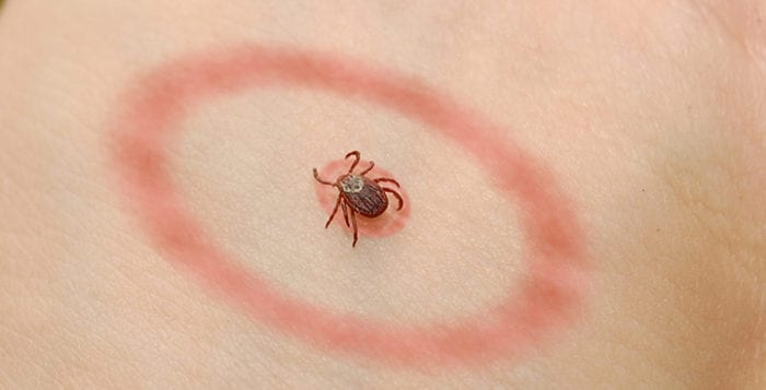
A plant-based diet involving lots of vegetables, fruits and some grains may have a similar effect as steroids in reducing inflammation. Stock photo
Steroids reduce inflammation, but it comes at a cost

By David Dunaief, M.D.

Dr. David Dunaief

Steroid use as a performance-enhancing drug was a significant factor in the recent Olympics, with the Russian team banned for their illegal use. However, if we look beyond the flashy headlines to rudimentary use, we see that corticosteroids, or steroids, play an important role in medicine.

This is a commonly prescribed class of medications. In fact, our bodies make corticosteroids, the indigenous form of steroids, in the cortex of the adrenals, glands that sit on top of the kidneys. Here, we are going to concentrate on the exogenous form, meaning from the outside as medication.

Use or benefit

Steroids have an anti-inflammatory effect. This is critical since many acute and chronic diseases are based at least partially on inflammation. Chronic diseases that benefit include allergic, inflammatory and immunological diseases (1). These types of diseases touch on almost every area of the body from osteoarthritis and autoimmune diseases — rheumatoid arthritis, inflammatory bowel disease, multiple sclerosis, lupus, psoriasis and eczema — to asthma, COPD (emphysema and chronic bronchitis) and eye disorders. This type of medication is pervasive.

Delivery

Steroids are delivered orally, topically as creams, lotions and eye drops, or via injections, intravenous solutions and inhaled formulations. The most commonly known medication is prednisone, but there is a plethora of others, including prednisolone, methylprednisolone, cortisone, hydrocortisone and dexamethasone. Their benefits can be tremendous, improving functionality and reducing pain or improving breathing. You could say they are lifesaving in some instances, and with rescue inhalers they may just be that.

The bad

However, there is a very big caveat: they come at a price. Steroids have lots of adverse events associated with them. This is where the bad part comes in and keeps on coming. Steroids cause weight gain, increased glucose (sugars), high blood pressure, cardiovascular events, osteoporosis, change in mood (psychoses), cataracts, glaucoma, infection, peptic ulcers, Cushing’s syndrome and the list goes on. Ironically, steroids help with breathing; however, as I’ve seen in my clinical experience, they can cause shortness of breath when weaned from a longer-use high dose too quickly.

The upshot

The good news is that a plant-based diet may have similar beneficial effects in chronic diseases as steroids without all the downsides. Let’s look at the evidence.

The role in pneumonia

Pneumonia is among the top-10 leading causes of death in the world (2). It can be a most painful and debilitating disease. I know, for I experienced it personally while I was in my medical training. Every time I coughed, it felt like there was a fire in my chest.

In a meta-analysis (a group of nine studies), there was no overall effect of corticosteroids in reducing the risk of mortality in community-acquired pneumonia (3). However, don’t fret; when the data was broken into subsets, the findings were different. In subset data of those who had severe pneumonia, there was a statistically significant 74 percent reduction in mortality. And when duration was the main focus in subgroup analysis, those who received prolonged use of steroids reduced their risk of mortality by half.

Unfortunately, with the benefit comes an increased risk of adverse events, and this meta-analysis was no exception. There was a greater than two-times increased risk of abnormally high glucose levels with prolonged use. Thus, when giving steroids, especially for a prolonged use, it may be wise to check glucose levels.

In a more recent randomized controlled trial (RCT), the gold standard of studies, the results reinforced the beneficial effects of steroids on pneumonia. They showed that in those with both severe pneumonia and high inflammation, there was a two-thirds reduction in treatment failures when corticosteroids were added to the regimen (4). There were 120 patients involved in the study. They received antibiotics plus either methylprednisolone or placebo for five days.

Osteoarthritis: surprising results

As we know, osteoarthritis specifically of the knee is very common, especially as the population continues to age. Intra-articular (in the joint) injections directly into the knee are becoming routine treatment. A recent study compared injectable hyaluronic acid to injectable corticosteroid (5). The results showed that over three months, the corticosteroid was superior to hyaluronic acid in terms of reducing pain, 66 percent versus 43.8 percent, respectively. Interestingly, over the longer term, 12 months, hyaluronic acid reduced the pain and maintained its effect significantly longer than the steroid, 33 percent versus a meager 8.2 percent, respectively.

Study groups received five injections of either steroid or of hyaluronic acid directly to the knee over a five-week period. Thus, steroids may not always be the most effective choice when it comes to pain reduction. Hyaluronic acid may have caused this beneficial effect by reducing inflammation, protecting cartilage and preventing cell death, according to the authors.

COPD: length may not matter

It is not unusual to treat COPD patients with oral steroids. But what is the proper duration? The treatment paradigm has been two weeks with 40 mg of corticosteroids daily. However, results in an RCT showed that five days with 40 mg of corticosteroid was noninferior (equivalent) to 14 days of the same dosage and frequency (6). About one-third of patients in each group experienced a COPD exacerbation within the six-month duration of the trial. The hope is that the shorter use of steroids will mean fewer side effects. There were over 600 patients in this trial. We have come a long way; prior to 1999, eight weeks of steroids was a more commonplace approach to treating acute COPD exacerbations.

Topical steroid risk

Even topical creams and lotions are not immune to risk. For example, potent topical creams and lotions placed around the orbit of the eye with prolonged use may negatively affect vision (7). However, the evidence is based mostly on case reporting, which is a low level of evidence.

Dietary effect

One of the great things about steroids is that they reduce inflammation, and we know that the basis of greater than 80 percent of chronic disease is inflammation. A plant-based diet involving lots of vegetables, fruits and some grains may have a similar effect as steroids. The effect of diet on chronic disease may be to modify the immune system and reduce inflammation (8).

The bioactive substances from plants thought to be involved in this process are predominantly the carotenoids and the flavonoids. Thus, those patients who respond even minimally to steroids are likely to respond to a plant-based diet in much the same beneficial way without the downsides of a significant number of side effects. Diet, unlike steroids, can be used for a long duration and a high intake, with a direct relationship to improving disease outcomes.

In conclusion, it is always better to treat with the lowest effective dose for the shortest effective period when it comes to steroids. The complications of these drugs are enumerable and must always be weighed against the benefits. Sometimes, other drugs may have more beneficial effects over the long term, such as hyaluronic acid injections for knee osteoarthritis. A plant-based diet, with anti-inflammatory properties similar to steroids, may be a useful alternative for chronic disease or may be used alongside these drugs, possibly reducing their dosage and duration.

References: (1) uptodate.com.(2) N Engl J Med. 1995;333(24):1618-24. (3) PLoS One. 2012;7(10):e47926. (4) JAMA. 2015;313(7):677-686. (5) Open Access Rheum 2015;7:9-18. (6) JAMA. 2013;309(21):2223-31. (7) Australas J Dermatol. Mar 5, 2015. (8) Int J Vitam Nutr Res. 2008 Dec;78(6):293-8.

Dr. Dunaief is a speaker, author and local lifestyle medicine physician focusing on the integration of medicine, nutrition, fitness and stress management.

by -
0 2149
If there were ever a reason needed for obese patients to lose weight, treating atrial fibrillation should be on the top of the list. Stock photo
Medications treat rate or rhythm or prevent strokes

By David Dunaief, M.D

Dr. David Dunaief

Atrial fibrillation is the most common arrhythmia, an abnormal or irregular heartbeat, found in the U.S. Unfortunately, it can be very complicated to treat. Though there are several options, including medications and invasive procedures, it mostly boils down to symptomatic treatment rather than treating or reversing underlying causes.

What is AFib? It is an electrical malfunction that affects the atria, the two upper chambers of the heart, causing them to beat “irregularly irregular.” This means there is no set pattern that affects the rhythm and potentially causes a rapid heart rate. The result of this may be insufficient blood supply throughout the body.

Complications that may occur can be severely debilitating, such as stroke or even death. AFib’s prevalence is expected to more than double by 2030 (1). Risk factors include age (the older we get, the higher the probability), obesity, high blood pressure, premature atrial contractions and diabetes.

AFib is not always symptomatic; however, when it is, symptoms include shortness of breath, chest discomfort, light-headedness, fatigue and confusion. This arrhythmia can be diagnosed by electrocardiogram, but more likely with a 24-hour Holter monitor. The difficulty in diagnosing AFib sometimes is that it can be intermittent.

There may be a better way to diagnose AFib. In a study, the Zio patch, worn for 14 days, was more likely to show arrhythmia than a 24-hour Holter monitor (2). The Zio patch is a waterproof adhesive patch on the chest, worn like a Band-Aid, with one ECG lead. While 50 percent of patients found the Holter monitor to be unobtrusive, almost all patients found the Zio patch comfortable.

There are two main types of AFib, paroxysmal and persistent. Paroxysmal is acute, or sudden, and lasts for less than seven days, usually less than 24 hours. It tends to occur with greater frequency over time, but comes and goes. Persistent AFib is when it continues past seven days (3). AFib is a progressive disease, meaning it only gets worse, especially without treatment.

Medications are meant to treat either the rate or rhythm or prevent strokes from occurring. Those that treat rate include beta blockers, like metoprolol, and calcium channel blockers, such as diltiazem (Cardizem). Examples of medications that treat rhythm are amiodarone and sotalol. Then there are anticoagulants that are meant to prevent stroke, such as warfarin and some newer medications, dabigatran (Pradaxa), rivaroxaban (Xarelto) and apixaban (Eliquis). The newer anticoagulants are easier to administer but may have higher bleeding risks, in some circumstances with no antidote.

There is also ablation, an invasive procedure that requires threading a catheter through an artery, usually the femoral artery located in the groin, to reach the heart. In one type of ablation, the inappropriate nodes firing in the walls of the atria are ablated, or destroyed, using radiofrequency. This procedure causes scarring of atrial tissue. When successful, patients may no longer need medication.

Premature atrial contractions

Premature atrial contractions, abnormal extra beats that occur in the atrium, may be a predictor of atrial fibrillation. In a study, PACs alone, when compared to the Framingham Heart Study AFib risk algorithm (a conglomeration of risk factors that excludes PACs) resulted in higher risk of AFib (4). When there were more than 32 abnormal beats/hour, there was a significantly greater risk of AFib after 15 years of PACs. When taken together, PACs and the Framingham model were able to predict AFib risk better at 10 years out as well. Also, when the number of PACs doubled overall in patients, there was a 17 percent increased risk of AFib.

The role of obesity

There is good news and bad news with obesity in regards to AFib. Let’s first talk about the bad news. In studies, those who are obese are at significantly increased risk. In the Framingham Heart Study, the risk of developing AFib was 52 percent greater in men who were obese and 46 percent greater in women who were obese when compared to those of normal weight (5). Obesity is defined as a body mass index >30 kg/m², and normal weight as a BMI <25 kg/m². There were over 5,000 participants in this study with a follow-up of 13 years. The Danish Diet, Cancer and Health Study reinforces these results by showing that obese men were at a greater than twofold increased risk of developing AFib, and obese women were at a twofold increased risk (6).

Now the good news: Weight loss may help reduce the frequency of AFib episodes. That’s right; weight loss could be a simple treatment for this very dangerous arrhythmia. In a randomized controlled trial, the gold standard of studies, those in the intervention group lost significantly more weight, 14 kg (32 pounds) versus 3.6 kg (eight pounds), and saw a significant reduction in atrial fibrillation severity score compared to those in the control group (7). There were 150 patients involved in the study.

An AFSS includes duration, severity and frequency of atrial fibrillation. All three components in the AFSS were reduced in the intervention group compared to the control group. There was a 692-minute decrease in the time spent in AFib over 12 months in the intervention arm, whereas there was a 419-minute increase in the time in AFib in the control group. These results are potentially very powerful; this is the first study to demonstrate that managing risk factors may actually help manage the disease.

Caffeine

According to a meta-analysis (a group of six population-based studies) done in China, caffeine does not increase, and may even decrease, the risk of AFib (8). The study did not reach statistical significance. The authors surmised that drinking coffee on a regular basis may be beneficial because caffeine has antifibrosis properties. Fibrosis is the occurrence of excess fibrous tissue, in this case, in the atria, which most likely will have deleterious effects. Atrial fibrosis could be a preliminary contributing step to AFib. Since these were population-based studies, only an association can be made with this discovery, rather than a hard and fast link. Still, this is a surprising result.

However, in those who already have AFib, it seems that caffeine may exacerbate the frequency of symptomatic occurrences, at least anecdotally. With my patients, when we reduce or discontinue substances that have caffeine, such as coffee, tea and chocolate, the number of episodes of AFib seems to decline. I have also heard similar stories from my colleagues and their patients. So, think twice before running out and getting a cup of coffee if you have AFib. What we really need are randomized controlled studies done in patients with AFib, comparing people who consume caffeine regularly to those who have decreased or discontinued the substance.

The bottom line is this: If there were ever a reason needed for obese patients to lose weight, treating atrial fibrillation should be on the top of the list, especially since it is such a dangerous disease with potentially severe complications.

References:

(1) Am J Cardiol. 2013 Oct. 15;112:1142-1147. (2) Am J Med. 2014 Jan.;127:95.e11-7. (3) Uptodate.com. (4) Ann Intern Med. 2013;159:721-728. (5) JAMA. 2004;292:2471-2477. (6) Am J Med. 2005;118:489-495. (7) JAMA. 2013;310:2050-2060. (8) Canadian J Cardiol online. 2014 Jan. 6.

Dr. Dunaief is a speaker, author and local lifestyle medicine physician focusing on the integration of medicine, nutrition, fitness and stress management. For further information, visit www.medicalcompassmd.com or consult your personal physician.