Tags Posts tagged with "evolution"

evolution

Aardvark. Pixabay photo

An international scientific project that compares the genomes of 240 living species of mammals has identified transposable elements (TEs) – genes that can change their position within a genome, creating or reversing mutations and thus altering a cell’s genetic identity – as a crucial area of study to help uncover the evolutionary process of mammals and to better understand biodiversity. Stony Brook University’s Liliana M. Dávalos is a collaborator in the analyses of TEs for the project. Two new papers, one published in the current issue of Science, and the other in Molecular Biology and Evolution, highlight the findings.

This graphic depicts the range of recently accumulated transposable elements (TEs) among sample mammals by proportion of their genome. Image credit: Osmanski et al. 2023 Science

The past 100 million years has caused mammals to adapt to virtually every environment on the planet. The Zoonomia Project, of which Dávalos is a scientific contributor, has cataloged the diversity in mammalian genomes by completing comparative genomic DNA sequences from the 240 species. The team, which consists of more than 150 scientists worldwide, published their multi-year comparative genome analysis in the Science paper.

Dávalos studies how biodiversity changes through time and what biological processes fuel biodiversity. She teamed up with David Ray and his lab at Texas Tech University to qualitatively analyze the dynamics of TEs.

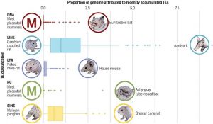
The paper describes the TE repertoires of 248 placental mammals. TEs make up a sizeable proportion of all mammalian genomes, yet there is much variation from one species to the next. The scientific team points out that that relating TEs to biodiversity is far from simple. Additionally, with the ability to move throughout the genome, TEs can contribute to biodiversity or also stymie it.

“Determining how many transposable elements of each kind are in each species is key to figuring out how transposons contribute to biodiversity. It seems simple to relate these counts to the number of species or their ecology, however that is misleading,” explains Dávalos, Professor of Conservation Biology in the Department of Ecology and Evolution, and a co-author of the paper. “Some species , like bats and whales, believe it or not are more closely related to each other than to others, such as bats and primates, so we must factor this related into our statistics within the comparative genomic mammalian analyses.”

The researchers identified more than 25,000 TE sequences in the mammalian set, with some mammals having large portions of TEs in their genome, calculated over time for each species. The average was approximately 45 percent

Overall, they concluded that “considering the wide-ranging effects that TEs impose on genomic architecture, these data are an important resource for future inquiries into mammalian genomics and evolution and suggest avenues for continued study of these important yet understudied genomic denizens.”

In the Molecular Biology and Evolution paper, novel statistical approaches to determining genome sequences in bats developed by Dávalos were used by the authors to describe the place bats hold with regard to TEs in mammals.

According to the lead author, Nicole Paulat, a graduate student in the Ray Lab, the research team found bats uniquely have more events involving TE transfers from one species to another. One mechanism that may explain such excess transfers is through viruses, an important finding on how several bat species have been found to host diverse and sometimes dangerous viruses.

Both papers based on the work from the Zoonomia Project illustrate that TEs are highly active across the genome of most mammal species, and because of this, future studies centering on TEs may help provide answers to mammalian biodiversity worldwide. Such research may also provide further hints as to how and why TEs disrupt mammalian genomes, therefore changing DNA and contributing to evolutionary processes and/or the development of disease.

The Brown Mouse Lemur (Microcebus rufus) is recognized as a vulnerable species on Madagascar. Photo by Chien C. Lee

A new study by a team of international scientists including Liliana M. Dávalos, PhD, of Stony Brook University’s Department of Ecology and Evolution, reveals that it would take three million years to recover the number of species that went extinct from human activity on Madagascar. Published in Nature Communications, the study also projects that if currently threatened species go extinct on Madagascar, recovering them would take more than 20 million years – much longer than what has previously been found on any other island archipelago in the world.

From unique baobab species to lemurs, the island of Madagascar is one of the world’s most important biodiversity hotspots. Approximately 90 percent of its species of plants and animals are found nowhere else. After humans settled on the island about 2,500 years ago, Madagascar experienced many extinctions, including giant lemurs, elephant birds and dwarf hippos.

Yet unlike most islands, Madagascar’s fauna is still relatively intact. Over two hundred species of mammals still survive on the island, including unique species such as the fossa and the ring-tailed lemur. Alarmingly, over half of these species are threatened with extinction, primarily from habitat transformation for agriculture. How much has human activity perturbed Madagascar away from its past state, and what is at stake if environmental change continues?

The team of biologists and paleontologists from Europe, Madagascar and the United States set out to answer this question by building an unprecedented new dataset describing the evolutionary relationships of all species of mammals that were present on Madagascar at the time that humans colonized the island.

As a co-author of “The macroevolutionary impact of recent and imminent mammal extinctions on Madagascar,” Daválos helped design the study, interpret a previously published lemur phylogeny, and analyzed prospects for new species discovery in Madagascar.

The dataset includes species that have already gone extinct and are only known from fossils, as well as all living species of Malagasy mammals. The researchers identified 249 species in total, 30 of which already are extinct. Over 120 of the 219 species of mammals that remain on the island today are currently classified as threatened with extinction by the IUCN Red List, due to habitat destruction, climate change and hunting.

Using a computer simulation model based on island biogeography theory, the team, led by Nathan Michielsen and Luis Valente from the University of Groningen (Netherlands) and Naturalis Biodiversity Center (Netherlands) found that it would take approximately three million years to regain the number of mammal species that were lost from Madagascar in the time since humans arrived.

The research team also determined through the computer simulation that if currently threatened species go extinct, it would take much longer: about 23 million years of evolution would be needed to recover the same number of species. Just in the last decade, this figure has increased by several million years, as human impact on the island continues to grow.

The amount of  time it would take to recover this mammalian diversity surprised the international team of scientists.

“These staggering results highlight the importance of effective conservation efforts in Madagascar. Here at Stony Brook, we can have an extraordinary impact on preventing extinction because of the longstanding biological field research at Centre ValBio and the associated Ranomafana National Park, with ongoing research on conservation while enhancing local livelihoods,” said Dávalos.

“It was already known that Madagascar was a hotspot of biodiversity, but this new research puts into context just how valuable this diversity is,” says leading researcher Luis Valente, Assistant Professor at the University of Groningen. “The time it would take to recover this diversity is much longer than what previous studies have found on other islands, such as New Zealand or in the Caribbean.”

The study findings ultimately suggest that an extinction wave with deep evolutionary impact is imminent on Madagascar, unless immediate conservation actions are taken. The good news – the computer simulation model shows that with adequate conservation action, we may still preserve over 20 million years of unique evolutionary history on the island.


Stock photo.

By Chris Zenyuh

Throughout our evolution, fruit stood as the primary source of sugars in our diet. That we evolved to desire sweetness, I contend, was not for energy but for the vitamins, minerals, fiber and antioxidants that come with the fruit. The fiber helps slow sugar absorption and reduce its negative metabolic potential, and the vitamins compensate.

The limitations of seasonal fruit accessibility made getting too much of these sugars infrequent, at most. Access to purified cane sugar was limited as well, due its tropical origins. The cost of growing and shipping cane sugar slowed its consumption, certainly for those of lesser means. Still, the demand for sugar steadily increased, a fact that the English monarchy used to fund its war chest.

William Duffy (in his book “Sugar Blues”) has suggested that the sugar machine was largely behind English colonization and enslavement through the 1800s. Duffy suggests that denying sugar’s responsibility for metabolic dysfunction dates back to Dr. Thomas Willis, private physician to King Charles II. Willis both discovered and named diabetes mellitus. Smart enough to recognize the illness and its sugar-related cause, Willis was also smart enough to name it after “honey” instead of sugar, perhaps to keep his job and his head!

Enjoying rations of sugar and rum, tens of thousands of the British sailors who guarded the sugar routes fell ill and died from scurvy. School children are taught that scurvy is a vitamin C deficiency, as it was discovered that the symptoms could be reversed with the addition of citrus to the rations. Sadly, this well-known story promotes the denial of the cause: too much sugar (and rum). Our food, medical and supplement industries continue to promote the use of fortification and vitamin supplements to “protect” against illnesses like scurvy, rather than incur financial loses that would result from curtailed consumption of sugars.

The spiraling decline of our general health gained momentum in 1973, when then Secretary of Agriculture Earl Butz instituted a 180 degree change in the farm subsidy program. Prior to 1973, farmers were directed by the government to curtail production to keep the supply and demand for corn in check. Sometimes, the farmers were instructed not to grow corn but were compensated for lost income. The restricted supplies kept corn prices high, making it too expensive to use high fructose corn syrup as a sweetener. Sugar cane, expensive due to its tropical origins, found itself in a limited range of food products.

The new program launched in 1973 rewarded corn farmers for producing as much corn as possible. Soon, the science to produce more corn, then the science to engineer additional uses for the extra corn became big businesses. High fructose corn syrup and cattle feed businesses were early beneficiaries of the new system. The ranchers and corn refiners lobbied to pay below cost for corn. Corn farmers would lose money, but, the new farm bills enabled the farmers to make up their losses (and more) by receiving the subsidies, funded by tax dollars. That made it cheaper to feed cattle corn than to feed them grass and cheaper to sweeten food with high fructose corn syrup (HFCS) than with sugar.

Americans were now able to purchase foods sweetened with HFCS and corn-fed meat at much cheaper prices than ever before. The cost, of course, does not include the medical expenses that may be incurred from chronic exposure to glucose and fructose, though.

The Sugar Association, still burdened with the expense of sugar cane’s tropical origins, has expanded its use of sugar beets to become price competitive in the caloric sweetener market. Farmed and processed in the continental United States, sugar beets are used to sweeten processed foods almost as cheaply as HFCS. If the ingredient label doesn’t specify cane sugar, it may very well be beet sugar. Of course, it is still sucrose.

Now you know why caloric sweeteners are omnipresent in our food system and how “food” can be available so cheap. You might want to reconsider the amount that you consume of what nature so frugally offers. Regardless of its source or history, it is metabolically the same!

Chris Zenyuh is a science teacher at Harborfields High School and has been teaching for
30 years.

'A trip to the American Museum of Natural History was my idea of being in heaven.' - Elof Carlson

By Elof Axel Carlson

The life sciences are vast in the number of specialties that exist for those pursuing a career as a biologist. A majority of college biology majors are premedical or seek some sort of health-related field. As much as possible they hope the biology they learn will find its way into the health field they seek to enter. Persons who want to be scholars in biology are often motivated by a desire to know as much about life as they can. I was one of those from early childhood when a trip to the American Museum of Natural History was my idea of being in heaven.

Elof Axel Carlson

I loved learning about evolution and the diversity of life. I knew I wanted to be a geneticist when I was in ninth grade and learned about Paul Müller’s Nobel Prize work on inducing mutations. Like a duckling, I felt imprinted and wanted to work with Müller someday.

Graduate work was different. As a teaching assistant I got to see about 90 different specimens each week for the various organ systems displayed by students. Unlike the textbook perfect illustrations, veins and arteries could be slightly off in the specimens I looked at. Their colors differed. Their texture differed.

I also learned how much we didn’t know about life. For my specialty of genetics (with Müller, as I had hoped) I felt steeped in experimental design, techniques and ways of thinking. Doing a Ph.D. allowed me to examine a gene using the tools of X-raying to produce mutations of a particular gene and subtle genetic design to combine pieces of a gene — taking it apart and combining pieces that were slightly different. It gave me an insight into that gene (dumpy, in fruit flies) that for a short time (until I published my work) I was the only person in the world that knew its structure.

In my career I have taught biology for majors, biology for nonscience majors, genetics, human genetics and the history of genetics. I have taught lower division and upper division courses, graduate courses and first-year medical classes. I learned that sharing new knowledge with students excited their imaginations. I learned that the human disorders I discussed led to office visits; and if I didn’t know the information they sought, I went with them to the medical library and we looked up articles in the Index Medicus and discussed their significance.

Often that student was married and had a child with a birth defect (born without a thyroid, having a family trait that might appear like cystic fibrosis). I would prepare a genetic pedigree and give it to the student to stick in a family bible for future generations to read. I also delighted in going to meetings to discuss genetics with colleagues whose work I had read.

I was pleased that I shared a body plan with other mammals. I liked comparative anatomy, which taught me how other body plans work (mollusks, arthropods, worms, coelenterates, echinoderms). As a graduate student taking a vertebrate biology course, I went into a cave and plucked hibernating bats from a ceiling.

The world under a microscope is very different. To see amoebas, ciliated protozoans, rotifers and other organisms invisible to the naked eye or as mere dust-like specks is a thrill. I can go back in time and imagine myself as a toddler, a newborn, an embryo in my mother’s uterus or an implanting blastocyst rolling out of her fallopian tube. I can imagine myself as a zygote, beginning my journey as a one-celled potential organism typing this article into a computer. I can go back in time to my prehistoric ancestors and trace my evolution back to the first cellular organism (bacteria-like) more than 3 billion years ago.

I learned, too, that I contain multitudes of ancestors who gave me one or more of their genes for the 20,000 I got from my father’s sperm and the matching 20,000 genes in my mother’s egg nucleus. I contain some 37 trillion (that is, 37,000,000,000,000) cells or 2 to the 45th power, which means some 45 mitotic cell divisions since I was a zygote. I know that the warmth of my body is largely a product of the mitochondrial organelles in my cells that using the oxygen from the air I breathe and converting small molecules of digested food to provide energy that runs the metabolism of my body and disposes carbon dioxide that eventually is expelled from my lungs. This knowledge makes me aware of my vulnerability at the cellular level, the chromosome level and the genetic level of my DNA to the agents around me that can lead to birth defects cancers, and a premature aging.

Knowing my biology allows me to know my risks as well as new ways to celebrate my life.

Elof Axel Carlson is a distinguished teaching professor emeritus in the Department of Biochemistry and Cell Biology at Stony Brook University.

Most snakes ... have no sign of limb development when X-rayed or when their skeletons are studied by anatomists.

By Elof Axel Carlson

Elof Carlson
Elof Carlson

The re-discovery of Mendelism in 1900 greatly enhanced breeding for new varieties of animals and plants. Similarly, the discovery of sex chromosomes and the chromosome theory of heredity enhanced Mendelism five years later. The discovery in the 1970s of genes controlling embryonic organs and body plans enhanced both embryology and genetics.

Also in the late 20th century a molecular approach was worked out that allows detection of genes and their functions using DNA sequencing and tools for isolating and inserting genes from one species into another. A good example of this is the analysis of limb development in vertebrates.

We are vertebrates because we have a spinal column and so do fish, frogs and tetrapods (four-limbed organisms like mice, humans, deer and lizards).

But some vertebrates lack limbs. Snakes are the best example of this. Boas and pythons do have internal vestigial hind limbs but totally lack any rudiments of limbs for their forelimbs. Most snakes, like vipers, have no sign of limb development when X-rayed or when their skeletons are studied by anatomists.

How did the snakes lose their limbs? The earliest ancestors of snakes did have hind limbs. Those ancestral types are only known from the fossil record. In the 1970s Hox genes, which determine development from the head to the tail, were found in vertebrates. The Hox gene for limb development is Hox C-6. It is regulated by another gene called sonic hedgehog or Shh. In vipers the Shh gene regulating Hox C-6 is mutated for both fore and hind limb production. In pythons it is nonfunctional for forelimbs but mutated with an aborted development preventing full growth of the hind limb buds.

Just this year, molecular biologists used the new techniques of gene removal and transfer (using a tool called CRSPR) and removed the Shh gene from a mouse fertilized egg. It resulted in a limbless baby mouse. When they put a python’s Shh gene in a mouse embryo whose Shh gene was removed, the resulting baby mouse had vestigial nubbins. When a mouse fertilized egg had its own Shh gene removed and replaced by that of a fish or human Shh gene, the baby mouse had perfectly normal limb development.

This work by Axel Visel and colleagues at Lawrence Biology Laboratories in Berkeley was published in the journal Cell. What makes science so attractive to scholars is its convincing logic, tested by experiments, to explore, confirm or rule out different interpretations of a puzzle. The puzzle of nature in this case is why snakes lost their limbs.

Those who see it in human terms (because we often use the ancient Greek dictum “Man is the measure of all things”) may invoke that the snake was punished for its role in corrupting humans. The scientist, however, likes to see things in more detail. There is comparative anatomy, the fossil record, experimental embryology, gene identification for function, sequencing for recognizing the gene, molecular tools for isolating the gene and experimental means of altering fertilized eggs to follow their fate.

Elof Axel Carlson is a distinguished teaching professor emeritus in the Dept.of Biochemistry and Cell Biology at Stony Brook University.