Tags Posts tagged with "Jun Wang"

Jun Wang

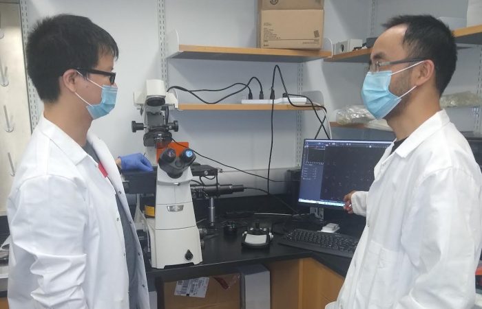
Study authors Liwei Yang, left, and Jun Wang, in the Wang laboratory by the microscope that incorporates the single-cell cyclic multiplex in situ tagging (CycMIST) technology to analyze proteins on single cells. Photo provided by Jun Wang

A new biomedical research tool that enables scientists to measure hundreds of functional proteins in a single cell could offer new insights into cell machinery. Led by Jun Wang, Associate Professor of Biomedical Engineering at Stony Brook University, this microchip assay — called the single-cell cyclic multiplex in situ tagging (CycMIST) technology – may help to advance fields such as molecular diagnostics and drug discovery. Details about the cyclic microchip assay method are published in  Nature Communications.

While newer technologies of single-cell omics (ie, genomics, transcriptomics, etc.) are revolutionizing the study of complex biological and cellular systems and scientists can analyze genome-wide sequences of individual cells, these technologies do not apply to proteins because they are not amplifiable like DNAs. Thus, protein analysis in single cells has not reached large-scale experimentation. Because proteins represent cell functions and biomarkers for cell types and disease diagnosis, further analysis on a single-cell basis is needed.

“The CycMIST assay enables comprehensive evaluation of cellular functions and physiological status by examining 100 times more protein types than conventional immunofluorescence staining, which is a distinctive feature not achievable by any other similar technology,” explains Liwei Yang, lead author of the study and a postdoctoral scholar within the Wang research team and Multiplex Biotechnology Laboratory.

Wang, who is affiliated with the Renaissance School of Medicine and Stony Brook Cancer Center, and colleagues demonstrated CycMIST by detecting 182 proteins that include surface markers, neuron function proteins, neurodegeneration markers, signaling pathway proteins and transcription factors. They used a model of Alzheimer’s Disease (AD) in mice to validate the technology and method.

By analyzing the 182 proteins with CycMIST, they were able to perform a functional protein analysis that revealed the deep heterogeneity of brain cells, distinguished AD markers, and identified AD pathogenesis mechanisms.

With this detailed way to unravel proteins in the AD model, the team suggests that such functional protein analysis could be promising for new drug targets for AD, for which there is not yet an effective treatment. And they provide a landscape of potential drug targets at the cellular level from the CycMIST protein analysis.

The authors believe that CycMIST could also have enormous potential for commercialization.

They say that before this study model with CycMIST, researchers could only measure and know a tip of protein types in a cell. But this new approach enables scientists to identify and know the actions of each aspect of a cell, and therefore they can potentially identify if a cell is in a disease status or not – the first step in a possible way to diagnose disease by analyzing a single protein cell. And compared with standard approaches like flow cytometry, their approach with CycMIST can analyze 10 times the amount of proteins and on a single-cell level.

The researchers also suggest that the cyclic microchip assay is portable, inexpensive, and could be adapted to any existing fluorescence microscope, which are additional reasons for its marketability if it proves to be effective with subsequent experimentation.

Much of the research for this study was supported by the National Institutes of Health’s National Institute of Aging (grant # R21AG072076), other NIH grants, and a Memorial Sloan Kettering Cancer Center Support Grant.

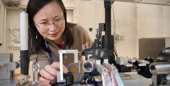
Jun Wang in her laboratory with a transmission x-ray microscope. Photo from BNL

By Daniel Dunaief

The first time is most definitely not the charm. That’s what Jun Wang and her colleagues at Brookhaven National Laboratory discovered about sodium ion batteries.

Wang, a physicist and lead scientist at the facility, looked deep into the inner workings of a sodium ion battery to determine what causes structural defects as the battery functions. As it turns out, the first time a sodium ion battery charges and discharges, it develops changes in the microstructure and chemical composition of iron sulfide. These changes, which degrade the performance of the battery, are irreversible during the first charging cycle.

“We found that the cracks happened during the first cycle, then, after that, the structure kind of reached equilibrium,” said Wang, who published her research in the journal Advanced Energy Materials. “All these changes happen during the first cycle.”

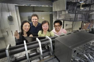
Collaborators from Brookhaven’s Photon Sciences and Sustainable Energy Technologies groups stand behind the new transmission x-ray microscope (TXM) at BNL’s National Synchrotron Light Source. From left: Yu-chen Karen Chen-Wiegart, Can Erdonmez, Jun Wang (team leader), and Christopher Eng. Photo from BNL

Sodium ion batteries are considered an alternative to lithium ion batteries, which are typically found in most consumer electronics. Like lithium, sodium is an alkali metal, which means that it is in the same group in the periodic table. Sodium, however, is more abundant and, as a result, considerably less expensive than lithium.

Using a synchrotron-based hard X-ray full-field microscope, Wang was able to see what happened when sodium ions moved into and out of an iron sulfide electrode through 10 cycles. “We can see this microstructure evolution,” she said.

Wang monitored the evolution as a function of time while the battery is charging and discharging. The results are the first time anyone has studied a sodium-metal sulfide battery with these tools, which provides information that isn’t available through other methods. “It is challenging to prepare a working sodium ion battery for the in operandi/in situ TXM study to correlate the microstructural evolution with its electrochemical performance,” she said.

Other researchers suggested that Wang has developed a following in the scientific community for her ground-breaking research. “She has a very good reputation in the area of X-ray nanotomography, applied to a wide range of different materials,” Scott Barnett, a professor of materials science and engineering at Northwestern University, explained in an email. “I am most familiar with her work on fuel cell and battery electrodes — I think it is fair to say that this work has been some of the best pioneering research in this area,” he said.

Barnett, who started collaborating with Wang in 2010 on measuring fuel cell and battery electrodes with X-ray tomography, suggested that Wang’s work on capacity loss “could certainly lead to new breakthroughs in improved batteries.”

In her most recent work with sodium ion batteries, Wang found that the defects start at the surface of the iron sulfide particles and move inward toward the core, Wang said. The microstructure changes during the first cycle and is more severe during sodiation. The particles don’t return to their original volume and shape. After the first cycle, the particles reach a structural equilibrium with no further significant morphological changes, she said.

In other cycles, the material does not show further significant morphological changes, reach a structural equilibrium and electrochemical reversibility. Wang and her colleagues confirmed these observations with X-ray nanotomography, which creates a three-dimensional image of the battery material while recording the change in volume.

Wang suggested that a way to reduce these structural defects could be to reduce the size of the iron sulfide particles to create a one-phase reaction. She will work with other collaborators on modeling and simulations that will enhance the design of future battery materials.

In addition to conducting research on batteries, Wang is an industrial program coordinator in the Photon Science Directorate at BNL. She works with industrial researchers and beamline staff to find and explore new opportunities in industrial applications using synchrotron radiation. She leads the industrial research program, interacting with user groups through consultation, collaboration and outreach.

To manage her research, which includes a lab of three other researchers, and to accomplish her mission as manager of an industrial research program, Wang jokes that she “spends 100 percent of her time” with each responsibility. “I try to do my best for the different things” she needs to do with her time, she said.

Jun Wang with her husband Qun Shen and their 11-year old son Sam in Waikiki last year. Photo from Jun Wang

A native of Wuhu, China, Wang earned her bachelor’s degree in physics from Anhui University in China and her doctorate in physics from the Chinese Academy of Sciences in Beijing. She worked at the Beijing Synchrotron Radiation Facility, which was the first synchrotron light source in China. During her doctoral training, she studied multilayer films using X-ray diffraction and scattering.

A resident of Poquott, Wang is married to Qun Shen, who is the deputy director for science at the NSLS-II. The couple has an 11-year-old son, Sam, who is a sixth-grade student at Setauket Elementary School. Shen and Wang met at an international X-ray crystallography conference in the early 1990s.

Shen trained in the United States after he graduated from Beijing University in 1980, when he went to Purdue University for his doctorate through the China-US Physica Examination and Application Program. The couple have worked together a few times over the years, including publishing a paper in Nature Communications. Wang is hoping that her work with battery research will lead to improvements in the manufacture and design of sodium ion batteries.