By Matthew Kearns, DVM
 February is National Pet Dental Health Month so I figured an article on dentistry is appropriate. A quick tooth anatomy reference: Everything below the gumline is considered the “root,” and everything above the gumline is considered the “crown.”
Feline odontoclastic resorptive lesions, or FORLs for short, is a strange pathology of the feline tooth, essentially a “hiccup” in the tooth loss cycle. Normal deciduous (baby) tooth loss involves the root and the structures around the root below the gumline. The destruction of the dentin is achieved by a type of cell called odontoclasts.
Odontoclasts are specialized cells whose sole job is to absorb the bone of the tooth root and the weakening of the structures around it. This process is critical in the loss of deciduous, or baby teeth, to make room for adult teeth to erupt.
I previously described FORLs as a hiccup in this normal tooth loss cycle because these same odontoclasts destroy the dentin, or layer of the tooth just below the enamel. The FORLs pathology progresses to invade the pulp below the dentin, and this is where the blood supply for the entire tooth lies. Once the blood supply is compromised, the enamel on the crown of the tooth starts to resorb, exposing the dentin underneath. This is above the gumline and can be painful as H-E-“double hockey stick.”
FORLs differ from a cavity where bacteria adhere to the surface of the crown of the tooth (the portion of the tooth above the gumline). Bacteria in the mouth produce acid that eats away the enamel until the root of the tooth is exposed.
Cavities are very unusual in dogs and cats for multiple reasons: the shape of the tooth, diet, type of bacteria in oral cavity compared to humans, and dogs and cats live shorter lives than humans. Dogs and cats suffer more from periodontal disease, or problems associated with structures around the tooth.
What triggers FORLs in cats? There are theories that include previous trauma, accumulation of plaque and tartar, impacted teeth around the affected tooth, etc. Bottom line, we as a veterinary community, do not yet know what triggers this pathology (to try to come up with a way to prevent it).
Treatment of FORLs requires removing the damaged portion of the tooth. This is a far less complicated and painful process compared to extraction, but the only way to determine if there is damage to the tooth is to perform dental X-rays. If the root is not involved, then it is FORLs (no extraction, just remove the damaged portion of the tooth); and if the root is involved, it is periodontal disease (extraction).
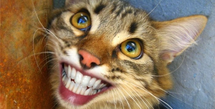
As more information becomes available about FORLs, I will give an update. Until then, make sure to get your cat (or dog) in for a dental checkup. SMILE!!!!!
Dr. Kearns practices veterinary medicine from his Port Jefferson office and is pictured with his son Matthew and his dog Jasmine.


