Tags Posts tagged with "Brain"

Brain

Sleep clears toxins from the brain. METRO photo
Exercise and sleep are crucial to clearing the clutter

By David Dunaief, M.D.

Dr. David Dunaief

Considering the importance of our brain to our functioning, it’s startling how little we know about it. 

We do know that certain drugs, head injuries and lifestyle choices negatively impact the brain. There are also numerous disorders and diseases that affect the brain, including neurological (dementia, Parkinson’s, stroke), infectious (meningitis), rheumatologic (lupus and rheumatoid arthritis), cancer (primary and secondary tumors), psychiatric mood disorders (depression, anxiety, schizophrenia), diabetes and heart disease.

Although these diseases vary widely, they generally have three signs and symptoms in common: they either cause altered mental status, physical weakness or change in mood — or a combination of these.

Probably our greatest fear regarding the brain is a loss of cognition. Fortunately, there are several studies that show we may be able to prevent cognitive decline by altering modifiable risk factors. They involve rather simple lifestyle changes: sleep, exercise and possibly omega-3s.

Let’s look at the evidence.

Clutter slows us down as we age

The lack of control over our mental capabilities as we age frightens many of us. Those who are in their 20s seem to be much sharper and quicker. But are they really?

In a study, German researchers found that educated older people tend to have a larger mental database of words and phrases to pull from since they have been around longer and have more experience (1). When this is factored into the equation, the difference in terms of age-related cognitive decline becomes negligible.

This study involved data mining and creating simulations. It showed that mental slowing may be at least partially related to the amount of clutter or data that we accumulate over the years. The more you know, the harder it becomes to come up with a simple answer to something. We may need a reboot just like a computer. This may be possible through sleep, exercise and omega-3s.

Get enough sleep

Why should we dedicate 33 percent of our lives to sleep? There are several good reasons. One involves clearing the mind, and another involves improving our economic outlook.

For the former, a study done in mice shows that sleep may help the brain remove waste, such as those all-too-dangerous beta-amyloid plaques (2). When we have excessive plaque buildup in the brain, it may be a sign of Alzheimer’s. When mice were sleeping, the interstitial space (the space between brain gyri, or structures) increased by as much as 60 percent.

This allowed the lymphatic system, with its cerebrospinal fluid, to clear out plaques, toxins and other waste that had developed during waking hours. With the enlargement of the interstitial space during sleep, waste removal was quicker and more thorough, because cerebrospinal fluid could reach much farther into the spaces. A similar effect was seen when the mice were anesthetized.

In another study, done in Australia, results showed that sleep deprivation may have been responsible for an almost one percent decline in gross domestic product for the country (3). The reason? People are not as productive at work when they don’t get enough sleep. They tend to be more irritable, and concentration may be affected. We may be able to turn on and off sleepiness on short-term basis, depending on the environment, but we can’t do this continually.

According to the Centers for Disease Control and Prevention, four percent of Americans reported having fallen asleep in the past 30 days behind the wheel of a car (4). And “drowsy driving” led to 83,000 crashes in a four-year period ending in 2009, according to The National Highway Traffic Safety Administration.

Prioritize exercise

How can I exercise, when I can’t even get enough sleep? Well there is a study that just may inspire you.

In the study, which involved rats, those that were not allowed to exercise were found to have rewired neurons in the area of their medulla, the part of the brain involved in breathing and other involuntary activities. There was more sympathetic (excitatory) stimulus that could lead to increased risk of heart disease (5). In rats allowed to exercise regularly, there was no unusual wiring, and sympathetic stimuli remained constant. This may imply that being sedentary has negative effects on both the brain and the heart.

This is intriguing since we used to think that our brain’s plasticity, or ability to grow and connect neurons, was finite and stopped after adolescence. This study’s implication is that a lack of exercise causes unwanted new connections. Human studies should be done to confirm this impact.

Consume omega-3 fatty acids

In the Women’s Health Initiative Memory Study of Magnetic Resonance Imaging Study, results showed that those postmenopausal women who were in the highest quartile of omega-3 fatty acids had significantly greater brain volume and hippocampal volume than those in the lowest quartile (6). The hippocampus is involved in memory and cognitive function.

Specifically, the researchers looked at the levels of eicosapentaenoic acid (EPA) and docosahexaenoic acid (DHA) in red blood cell membranes. The source of the omega-3 fatty acids could either have been from fish or supplementation.

It’s never too late to improve brain function. Although we have a lot to learn about the functioning of the brain, we know that there are relatively simple ways we can positively influence it.

References:

(1) Top Cogn Sci. 2014 Jan.;6:5-42. (2) Science. 2013 Oct. 18;342:373-377. (3) Sleep. 2006 Mar.;29:299-305. (4) cdc.gov. (5) J Comp Neurol. 2014 Feb. 15;522:499-513. (6) Neurology. 2014;82:435-442.

Dr. David Dunaief is a speaker, author and local lifestyle medicine physician focusing on the integration of medicine, nutrition, fitness and stress management. For further information, visit www.medicalcompassmd.com. 

by -
0 1276

There is a lot of stress in our lives these days. Stress envelops us. One man I know complained that even in his home, he does not feel stress free. When he puts on the television or radio, the now-commonplace partisan viewpoints surround him. And that is the least of it. The horrific shooting in a Pittsburgh synagogue, pipe bombs sent to at least 15 different targets perceived to be Democratic in nature throughout the United States, the shooting at a school in North Carolina and more make up some of the news just this past week. There seems to be no escape. Even conversation with customers or spouses inevitably touches on the daily stressful events.

Surely there have been times of even greater stress in our country. World War II comes immediately to mind. The Cold War, with regular air raids, was another. The Cuban Missile Crisis was yet another. But these were all threats from outside: from the Nazis, the Japanese, the Soviet Union. The stress today, whether rhetorical or physical, is domestic and aimed by Americans against other Americans. Worst of all, as political partisans denigrate opponents and gun violence becomes tragically routine, there doesn’t seem to be an end in sight.

Can we learn to manage the stress in our lives? The Harvard Women’s Health Watch advises that we can. In the August issue, published by a division of Harvard Medical School, physicians offer some information about stress and its effects. They also give some suggestions for coping with stress.

First the information. “It’s not uncommon to feel disorganized and forgetful when you’re under a lot of stress,” the article, “Protect your brain from stress,” explained. “But over the long term, stress may actually change your brain in ways that affect your memory.” Because stress can influence how the brain functions, including not only memory but also mood and anxiety, it can cause inflammation. This in turn can affect heart health. Thus stress has been associated with multiple chronic diseases of the brain and heart, according to Harvard physicians.

The brain is not just a single unit but a group of different parts that perform different tasks, according to the Harvard article. When one part is engaged, researchers believe that other parts may not have as much energy for their specialized functions. One example is if you are in a dangerous situation, the amygdala section takes over to ensure survival, while the energy level in parts having to do with memory or higher-order tasks recedes. Hence you might be more forgetful when stressed.

“There is evidence that chronic (persistent) stress may actually rewire your brain,” according to the research, as if exercising one section makes it stronger while other sections, like that having to do with more complex thought, take “a back seat.” Such brain changes may be reversible.

There are various kinds of stress. For example, one feels differently before taking a big test compared with that experienced in a car accident. More stress is worse, and long-term stress is generally worse than short-term stress, according to the physicians. Unpredictable stress is worse than stress that can be anticipated. Chronic stress can be more challenging than one that will end shortly. Feeling supported by others most likely mitigates stress effects.

So here is some advice from the Harvard publication on how to cope with stress. Establish some control over your situation such as by setting a routine. Get organized. Get a good night’s sleep — hard to do when stressed but going to bed and waking up at the same time each day helps, as does avoiding caffeine and creating a relaxing sleep environment. Get help, sooner rather than later. And try to change your attitude toward stress by striving for healthier responses to stress. Use its effects, if you can, to high power you to a goal. Like voting.

And I say, turn off the television and the instant news briefs on your cellphone for some quiet time each day.

We should dedicate 33 percent of our lives to sleep to improve brain health. Stock photo

By David Dunaief, M.D.

The brain is the most important and complex organ, yet what we know about the brain is inverse to its prominence. In other words, our knowledge only scratches the surface. While other organs can be transplanted readily, it is the one organ that can’t, at least not yet.

The brain also has something called the blood-brain barrier. This is an added layer of small, densely packed cells, or capillaries, that filter what substances from the blood they allow to pass through from the rest of the body (1). This is good, since it protects the brain from foreign substances; however, on the downside, it also makes it harder to treat, because many drugs and procedures have difficulty penetrating the blood-brain barrier.

Unfortunately, there are many things that negatively impact the brain, including certain drugs, head injuries and lifestyle choices. There are also numerous disorders and diseases that affect the brain, including neurological (dementia, Parkinson’s, stroke), infectious (meningitis), rheumatologic (lupus and rheumatoid arthritis), cancer (primary and secondary tumors), psychiatric mood disorders (depression, anxiety, schizophrenia), diabetes and heart disease.

These varied diseases tend to have three signs and symptoms in common: they either cause an alteration in mental status — cognitive decline, weakness or change in mood – or a combination of these.

Probably our greatest fear regarding the brain is cognitive decline. We have to ask ourselves if we are predestined to this decline, either because of the aging process alone or because of a family history, or if there is a third option, a way to alter this course. Dementia, whether mild or full-blown Alzheimer’s, is cruel; it robs us of functioning. We should be concerned about Alzheimer’s because 5.2 million Americans have the disease, and it is on the rise, especially since the population is aging (2).

Fortunately, there are several studies that show we may be able to choose the third option and prevent cognitive decline by altering modifiable risk factors. They involve rather simple lifestyle changes: sleep, exercise and possibly omega-3s. Let’s look at the evidence.

The impact of clutter

The lack of control over our mental capabilities as we age is what frightens us the most since we see friends, colleagues and relatives negatively affected by it. Those who are in their 20s seem to be much sharper and quicker. But are they really?

In a recent study, German researchers found that educated older people tend to have a larger mental database of words and phrases to pull from since they have been around longer and have more experience (3). When this is factored into the equation, the difference in terms of age-related cognitive decline becomes negligible. This study involved data mining and creating simulations. It showed that mental slowing may be at least partially related to the amount of clutter or data that we accumulate over the years. The more you know, the harder it becomes to come up with a simple answer to something. We may need a reboot just like a computer. This may be possible through sleep and exercise and omega-3s.

Sleep

I have heard people argue that sleep gets in the way of life. Why should we have to dedicate 33 percent of our lives to sleep? There are several good reasons. One involves clearing the mind, and another involves improving our economic outlook.

For the former, a study shows that sleep may help the brain remove waste, such as those all-too-dangerous beta-amyloid plaques (4). When we have excessive plaque buildup in the brain, it may be a sign of Alzheimer’s. This study was done in mice. When mice were sleeping, the interstitial space (the space between brain gyri, or structures) would increase by as much as 60 percent.

This allowed the lymphatic system, with its cerebrospinal fluid, to clear out plaques, toxins and other waste that had developed during waking hours. With the enlargement of the interstitial space during sleep, waste removal was quicker and more thorough because cerebrospinal fluid could reach much further into the spaces. When the mice were anesthetized, a similar effect was seen as with sleeping.

In a published follow-up study, the authors found that sleep position had an impact on glymphatic transport in rodents. Sleeping in a lateral position, or on their sides, was more effective at clearing waste than prone or supine positions. Of course, the authors note that for rodents a prone position is similar to their awake positions. It would be most like a human sleeping while sitting upright (5).

In another study, done in Australia, results showed that sleep deprivation may have been responsible for an almost 1 percent decline in gross domestic product for the country (6). The reason is obvious: People are not as productive at work when they don’t get enough sleep. Their attitude tends to be more irritable, and concentration may be affected. We may be able to turn on and off sleepiness on an acute, or short-term, basis, depending on the environment, but it’s not as if we can do this continually.

According to the Centers for Disease Control and Prevention, an average of 4 percent of Americans report having fallen asleep in the past month behind the wheel of a car (7). I hope this hammers home the importance of sleep.

Exercise

How can I exercise, when I can’t even get enough sleep? Well there is a study that just may inspire you to exercise.

In the study, which involved rats, those that were not allowed to exercise were found to have rewired neurons in the area of their medulla, the part of the brain involved in breathing and other involuntary activities. There was more sympathetic (excitatory) stimulus that could lead to increased risk of heart disease (8). In those rats that were allowed to exercise regularly, there was no unusual wiring, and sympathetic stimuli remained constant. This may imply that being sedentary has negative effects on both the brain and the heart.

This is intriguing since we used to think that our brain’s plasticity, or ability to grow and connect neurons, was finite and stopped after adolescence. This study’s implication is that a lack of exercise causes unwanted new connections. Of course, these results were done in rats and need to be studied in humans before we can make any definitive suggestions.

Omega-3 fatty acids

In the Women’s Health Initiative Memory Study of Magnetic Resonance Imaging Study, results showed that those postmenopausal women who were in the highest quartile of omega-3 fatty acids had significantly greater brain volume and hippocampal volume than those in the lowest quartile (9). The hippocampus is involved in memory and cognitive function.

Specifically, the researchers looked at the level of omega-3 fatty acids, called eicosapentaenoic acid and docosahexaenoic acid, in red blood cell membranes. The source of the omega-3 fatty acids could either have been from fish or supplementation. This was not delineated. The researchers suggest eating fish high in these substances, such as salmon and sardines, since it may not even be the omega-3s that are playing a role but some other substances in the fish. It’s never too late to improve brain function. You can still be sharp at a ripe old age. Although we have a lot to learn about the functioning of the brain, we know that there are relatively simple ways we can positively influence it.

References: (1) medicinenet.com. (2) alz.org. (3) Top Cogn Sci. 2014 Jan.;6:5-42. (4) Science. 2013 Oct. 18;342:373-377. (5) J Neurosci. 2015 Aug 5;35(31):11034-11044. (6) Sleep. 2006 Mar.;29:299-305. (7) cdc.gov. (8)J Comp Neurol. 2014 Feb. 15;522:499-513. (9) Neurology. 2014;82:435-442. Dr. Dunaief is a speaker, author and local lifestyle medicine physician focusing on the integration of medicine, nutrition, fitness and stress management.

When they work as they should, they become a part of a process that helps us remember the Amendments to the Constitution, the Pythagorean Theorem, or the words to a love poem by Elizabeth Barrett Browning. When they don’t work correctly, we can run into all kinds of problems, some of which can get worse over time.

The N-methyl-D-aspartate receptor, also known as the NMDA receptor, which has parts that are bound in the membrane of brain cells, or neurons, is at the center of learning and memory.

Up until last year, only parts of the NMDA receptors sticking out of the membrane were known. A lack of a three-dimensional understanding made it difficult to see how this receptor works. Hiro Furukawa, an associate professor at Cold Spring Harbor Laboratory, and his postdoctoral researcher, Erkan Karakas, provided considerably more structural details of this receptor.

“The structures of the full-length NMDA receptor that [Furukawa’s] lab generated last year are seminal,” said Lonnie Wollmuth, a professor in the Department of Neurobiology and Behavior at Stony Brook University and a collaborator with Furukawa on other work. “They are fundamental to understanding how the NMDA receptor operates and how it can be modified in the clinic.”

Wollmuth suggested Furukawa has an “outstanding” reputation and said the structure of the receptor will “drive the field in new directions.”

Furukawa cautioned that scientists are still missing a structural understanding of a piece of the receptor that protrudes into the cell. Seeing the structure of this receptor will “provide clues for developing new compounds and for redesigning existing compounds to minimize side effects associated with nonspecific targeting,” Furukawa explained.

When NMDA receptors open, sodium and calcium ions flow into the cells. Too much calcium in the cells can cause toxicity that results in the neurodegeneration observed in Alzheimer’s disease and injuries related to strokes. Changes in the concentration of these ions can excite the neuron and cause symptoms such as epilepsy.

Seeing the structure of this receptor can provide a road map to find places on it that can become too active or inactive. Researchers typically look for binding sites, where they can send in a drug that can affect the function of the receptor. The more binding pockets scientists like Furukawa find, the greater the opportunity to regulate the NMDA receptor function.

Furukawa’s lab includes two graduate students, four postdocs and a technician. He is collaborating with scientists at Emory University to design and synthesize novel compounds based on the protein structures. As he gets more research funding, Furukawa would like to add more expertise in bioinformatics, which involves using computer science and statistics to understand and interpret large collections of data.

Experts in this field can go through a database of compounds quickly, enabling scientists to conduct the equivalent of thousands of virtual experiments and screen out candidates that, for one reason or another, wouldn’t likely work.

Furukawa is also studying autoimmune disorders in which immune cells attack these important receptors. One of these diseases is called anti-NMDA receptor encephalitis. Susannah Cahalan wrote an autobiographical account of her struggle with the disease in a New York Times Best Selling Book called “Brain on Fire: My Month of Madness” in 2012.

Furukawa is collaborating with a group at the University of Pennsylvania to find a way to detect the autoimmune antibodies that causes encephalitis. He is working to find a way to quench autoimmune antibodies for an anti-NMDA receptor.

Furukawa lives in Cold Spring Harbor with his wife, Megumi, who used to be an elementary school teacher but is now taking care of their sons Ryoma, 7, and Rin, 4.

Furukawa, who moved from Japan to Boston in fifth grade, then back to Japan for junior high school and finished high school in Missouri, is enjoying an opportunity to grow his own vegetables on Long Island.

As an undergraduate at Tufts, Furukawa was more interested in international politics and economics than in science. When he took chemistry and physics classes, he said the work “clicked comfortably” and he wound up majoring in chemistry. As an eight-year-old, he recalled watching the stars at night through a telescope. When he saw a ring of Saturn for the first time, he was so excited that he couldn’t sleep.

Furukawa’s colleagues appreciate his dedication to his work.

“He is certainly driven,” said Wollmuth. “He is in an extremely competitive field, so he must work efficiently and hard.”

by -
0 2087
Professor Helen Benveniste. File photo from SBU

Sleeping doesn’t just provide a break from the daily grind, prevent you from chowing down on more Oreo cookies, or keep you out of trouble when it gets dark. It may also serve an important brain-cleaning function, getting rid of tau and beta amyloid proteins.

Merely shutting your eyes and letting the sandman sprinkle dust on your forehead may not be enough. You might actually help your brain, over the long term, with the way you sleep.

Helene Benveniste, a professor of anesthesiology and radiology and vice chair for research in the Department of Anesthesiology at Stony Brook University, recently conducted research on anesthetized rodents, tracking how the glymphatic system worked in various sleep positions. The animals were better at flushing tau and beta amyloid proteins from their brains when they slept laterally, or on their sides, than when they slept on their stomachs. Resting on their backs wasn’t as efficient as sleeping on their sides, although it was better than face down.

These proteins aren’t just a part of everyday maintenance. They likely play a role in the onset of Alzheimer’s disease and other age-related neurological problems, Benveniste said.

Since Benveniste published her study in the Journal of Neuroscience in early August, she has received a flood of emails from around the world, including from Brazil, France and Colombia, with people asking about various sleep positions and neurological disorders.

The Stony Brook professor said it is too soon after this study to come to any conclusions about sleep or preventing cognitive disorders. For starters, she and a research team that included scientists at the University of Rochester, NYU Langone Medical Center and Stony Brook conducted the studies on animal models, rather than on humans.

“In general, the rodent is a pretty good model for core aspects of human brain function,” said Dennis Choi, the chairman of the Neurology Department at Stony Brook. The specifics, however, can differ from one species to another. As a result, Benveniste said, “I don’t think anybody should panic” about the way he or she sleeps.

Scientists know that in the glymphatic pathway, cerebrospinal fluid moves through the brain and exchanges with interstitial fluid to get rid of waste. In the studies with rodents, the face down position seemed to divert the cerebrospinal fluid away from the brain, Benveniste said.

The research could be another step toward understanding how sleep might help with the human glymphatic system.

An anesthesiologist who does clinical work one day a week, Benveniste said she started thinking about conducting this kind of study a few years ago. Benveniste is a “good example of a physician/scientist,” Choi said.

Two years ago, a study by a co-author on the paper, Maiken Nedergaard from the University of Rochester, showed that sleep or general anesthesia enhances the clearance of waste from the brain of rodents.

“Since I am an anesthesiologist, I immediately thought about how body/head positions during anesthesia might affect clearance,” Benveniste said. The data took over a year and a half to collect and analyze.

“The quantitative aspect of this system should not be overlooked. To find out how these [proteins] are moving through the brain is a huge issue,” she said. The collaboration with Jean Logan, senior research scientist in the Department of Radiology at NYU “enabled us to move forward.”

Benveniste used a dynamic contrast MRI method to calculate the exchange rates between the cerebrospinal fluid and the interstitial fluid. The next step in these studies is to move toward the human brain. Benveniste said she is working with colleagues at the National Institutes of Health.

Just from observing wildlife outside the lab, Benveniste said many animals tend to sleep in what she and her team found was the optimum position for clearing waste in rodents: on their sides. “Even elephants lie down in recumbent, lateral positions,” she added.

As for Benveniste, she said she naturally sleeps on her right side. She said she’s well aware of how well she slept during the night. If she wakes up after getting enough rest, she said she thinks, “this was a good night’s sleep. This was good for my brain.”

Benveniste, who lives in Northport with her husband, Peter Huttemeier, is also an advocate of exercise for brain health, although she doesn’t suggest marathon running. “I do think this may be affecting the cerebrospinal fluid flow dynamics,” she said, adding that she wants to take up yoga.

Benveniste is eager to continue to build on this sleep study. “The workings of this system so far has been an amazing exploratory adventure,” she said.

Huntington neurosurgeon touts procedure’s success

This diffusion tensor imaging shows the patient’s nerve, brain and other tissue matter surrounding the white mass, which makes it easier for surgeons to diagnose and treat their patients. Photo from Ericca Ardito

When Jean Noschese’s left hand started to go numb, she didn’t expect her doctor’s visit to lead to brain surgery at Huntington Hospital, where she met Dr. Robert Kerr, a neurosurgeon who had a new way of operating on the brain.

On Oct. 16, 2013, Noschese experienced a head-on collision while driving in Hauppauge. The car accident left her in need of several surgeries, including ones to repair her rotator cuff and replace her hip. But it was when she started losing sensation in her left hand, in 2014, that she went to a specialist. Noschese, who initially wondered if her issue with her hand was related to her crash, was rushed to the hospital after her hand specialist thought Noschese was experiencing a stroke.

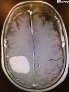
But instead of a stroke, the doctors found a three-by-four-centimeter lesion on the right side of her brain that caused paralysis on the left side of her body. Her lesion wasn’t caused by the crash, but from Noschese’s breast cancer that had metastasized to her brain. Noschese was diagnosed with breast cancer in 2005.

Doctors wanted to perform brain surgery the following day, on Dec. 22, 2014, according to Noschese,

“It’s overwhelming to hear that you need brain surgery,” she said.

A large mass is located in the left hemisphere of the brain prior to the operation where Dr. Robert Kerr used Synaptive Medical’s BrightMatter Plan. Photo from Ericca Ardito

But Kerr, who met with Noschese when she entered the hospital, reassured her and reviewed the procedure with her. A new brain-mapping technique, using the Synaptive Medical’s BrightMatter Plan product, involves several new tools, including a highly engineered tube that splits brain tissue fibers and allows neurosurgeons to access difficult and deep parts of the brain easily. The procedure also utilizes a fiber optic, high definition telescope that creates a “cone of visualization” that allows surgeons to clearly view all planes of the brain they are working on.

The technique also features a procedure called the myriad, which uses a blunt suction device that peels off tumors from dangerous or sensitive areas without damaging surrounding areas in the brain.

“Traditionally, surgeries for deeper regions actually involve destroying a certain amount of tissue to get to the target area,” Kerr said.

According to Kerr, in traditional brain surgeries, metal retractors are used to create a pathway so surgeons can access target areas of the brain. Doctors use the retractors to pull the edges of the brain apart and create a pathway.

Kerr said the issue with this technique is that, regardless of how careful a surgeon is, he or she may still push on these retractors, which widens the pathway the surgeon created from surface of the brain to the target area. As a result, the patient is left with a hole in part of the brain, which means the patient will take longer to recover from the surgery.

Stony Brook Medicine Neurosurgeon Dr. David Chesler said Synaptive Medical’s BrightMatter Plan procedure is only appropriate under certain circumstances.

“Tumors that come right to the surface, where they’re easily approachable, I don’t think there’s any benefit to using this technique, because the tumor is right there,” Chesler said. Chesler took a course for the procedure about two years ago. While he thinks the technique is beneficial, he does not think it is a be-all and end-all procedure for brain surgery.

While the procedure is minimally invasive, may decrease the chance of injuring the patient during the operation and allows surgeons to approach lesions or blood clots, Chesler said there are some downsides to the technique. He said that the technology of this technique is not new, but simply creates a new system that makes it easier for surgeons to implement.

Additionally, minimally invasive procedures double or triple the length of an operation, depending on the surgeon and the nature of the surgery. Surgeons who may not be very experienced may leave parts of lesions or tumors and blood clots behind because of limited visualization.

A large mass is located in the left hemisphere of the brain has been removed with the use of Synaptive Medical’s BrightMatter Plan. Photo from Ericca Ardito

Surgeons can only see what is at the end of the tube, which is around the diameter of a dime.

Kerr said this procedure will decrease patients’ recovery time. Patients are usually tired following the surgery and require extensive amounts of rest before they are discharged from the hospital. Noschese, however, was alert and speaking two hours after the surgery. Not only did she regain sensation in her hand, but she was also able to grab and hold onto a variety of objects.

Chesler, like Kerr, said patients who receive traditional surgeries for deep-seated lesions or blood clots can do well. He said his patients’ deficits were more related to the “structures involved with the tumor,” as opposed to the approach used. Chesler has seen both good and bad outcomes from this technique

According to Kerr, few surgeons are using this technique.

“Neurosurgeons are skeptics and slow adopters and I think that’s appropriate,” Kerr said, explaining why more surgeons may stick to traditional brain surgery practices.

While Chesler said the procedure should be used for the right case and with an experienced surgeon, he said staff are looking to adopt this technique at Stony Brook University Hospital. Chesler, who does both pediatric and adult neurosurgery, said he is simply looking for the right case. Recently he hasn’t come across a case that calls for the technique.

Doctors must demonstrate the procedure and illustrate its benefits when introducing the technique to the hospital. Hospitals need to invest in the procedure for a surgeon to officially implement it.

Although Chesler said there are other systems that surgeons can use to reach a similar goal, Kerr said the technique is a glimpse into the future of this type of surgery.

“I think this technology reflects the future of neurosurgery and accessing deep-seated lesions in a kinder, gentler, more precise way,” Kerr said. “I think this is representing a future paradigm shift in the way that brain surgery is done, and I think that we will see many more adopting this in a very short period of time.”