Ask the Vet: Hey Doc, do I need to worry about these lumps on my pet?
By Matthew Kearns, DVM

Working in a general practice for many years now, I am commonly asked the question of whether or not a growth under the skin is something to worry about or not. The good news is the majority of subcutaneous (under the skin) lumps are benign, or non-cancerous. They are usually lipomas (benign tumors made up of fat cells), or cysts. Some, however, can be cancerous and are better off being removed before they get too big or invade the surrounding tissues making it more difficult or impossible to remove completely. How are we to tell?
It is always good to bring any new growths to your veterinarian’s attention as soon as possible. When I evaluate any new subcutaneous growths I was taught to look for three characteristics:
Is it hard or is it soft? A growth that is hard is not always malignant, and a growth that is soft is not always benign. However, in my book, a growth that is hard is more concerning than a growth that is soft.
Is it freely movable or well attached to underlying tissues? Growths that are soft and freely movable are more likely to be benign than growths that are hard and well attached.
Is it painful, or non-painful? subcutaneous growths that are non-painful to palpation are less likely to be malignant.
I like to have the three criteria in combination to feel confident in telling a pet owner that the growth is something just to be monitored. Therefore soft, freely movable, non-painful is good, and firm, well-attached, and painful is concerning.
I also like to recommend owners monitor these growths closely for change. The two changes I look for is a rapid change in size, and/or a change in character. If a growth doubles in size in a month or less, or a change in character (soft to hard, freely movable to well attached, non-painful to painful) then please have your pet seen again immediately.
There are different ways to test these growths. My favorite first test is a fine needle aspirate and cytology. This test involves inserting a needle into the growth and aspirating a sample of cells. The sample is then sprayed onto a microscope slide, allowed to dry and sent to the lab for evaluation by a pathologist.
Cytology is the evaluation of individual cells so it is not as accurate as a biopsy but one can get a lot of information from individual cells. A fine needle aspirate is something that is well tolerated by most patients so sedation or anesthesia is not usually required to perform this procedure.
There are cases where the fine needle aspirate and cytology are inconclusive and a biopsy is needed. However, I feel a fine needle aspirate and cytology is an excellent first step. I hope this information helps.
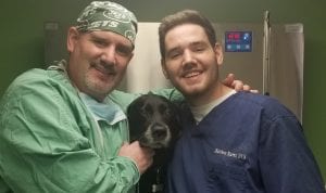
Dr. Kearns practices veterinary medicine from his Port Jefferson office and is pictured with his son Matthew and his dog Jasmine.


