
By Matthew Kearns, DVM
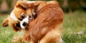
“My dog keeps licking at one area. Why does she do it?!!!!” The answer is quite simple. However, the diagnosis and treatment is quite complicated (and often quite frustrating). The answer is something called an acral lick granuloma.
Acral lick granulomas form when a dog repetitively licks at a spot (usually on one of the front legs) until a raised, inflamed, firm, hairless nodular growth on the skin. Breeds that are considered more at risk are Doberman Pinscher, Labrador Retriever, Golden Retriever, Great Dane, boxer, and Irish Setter.
Acral lick granulomas are multi-factorial, meaning many factors cause this condition. Additionally, acral lick granulomas usually have a primary cause and secondary complications. Primary causes include allergies (most common), trauma to the area, arthritis, skin parasites, deep fungal infections, tumors and behavioral issues.
Most veterinarians (including myself) will treat these conditions initially empirically. What this means is we will treat for the symptoms without investigating a cause. Treating empirically is a less expensive way (this keeps pet owners happy in my experience) to proceed and works in some cases. When it doesn’t, then a diagnostic workup is indicated (this does not keep pet owners happy in my experience).
Testing includes X-rays, bloodwork, cultures, and biopsies. Diagnosis of allergies (both food and environmental) is very important to either rule in, or rule out as part of the workup. This can include changing your dog’s diet, bloodwork, or even skin allergy testing.
Treatment for acral lick granulomas includes management of both the itch/pain, as well as the infection. Breaking the “itch-lick” cycle is very important. A combination of corticosteroids (cortisone derivatives) and antibiotics can be quite effective and is used initially by many veterinarians to see if they can resolve the problem without a large diagnostic workup.
Topical medications can be quite effective if the patient does not lick it off. Some sort of covering like a sock or bandage (if the patient will not pull off or eat) or an Elizabethan collar to keep the patient from licking at the area is often used with medication to break the cycle.
If a specific type of infection, whether it be fungal or bacterial, long-term antibiotics or antifungals may be needed. Also realize that even if your dog leaves the granuloma for long periods of time, flare ups are possible which requires treatment again.
Acral lick granulomas have a unique behavioral component to them. Dogs that have lick granulomas many times have other compulsive disorders or separation anxiety. Medications such as tricyclic antidepressants (TCAs) and selected serotonin reuptake inhibitors (SSRIs) are used in conjunction with other medications to break the “itch-lick” cycle in compulsive patients.
If your veterinarian makes a diagnosis or tentative diagnosis of an acral lick granuloma be patient with your dog and your veterinarian.
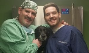
Dr. Kearns practices veterinary medicine from his Port Jefferson office and is pictured with his son Matthew and his dog Jasmine.


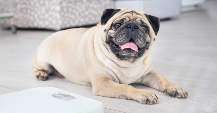


 Arthritis is a condition that will be exacerbated by cold weather just the same as humans. Arthritis is also complicated by weight gain and weight gain is common in pets in cold winters due to inactivity. Consider giving a little less food and be very judicious with treats (COVID has fattened up some pets at our practice with owners working from home). Pets with arthritis are more likely to slip on snow or ice so make sure to clear a path for them when they go out and assist them if necessary. Joint supplements are excellent year-round but, if you have forgotten to continue through the winter we recommend restarting immediately.


